Arteriole smooth muscle cells, light micrograph
Bildnummer 12360919
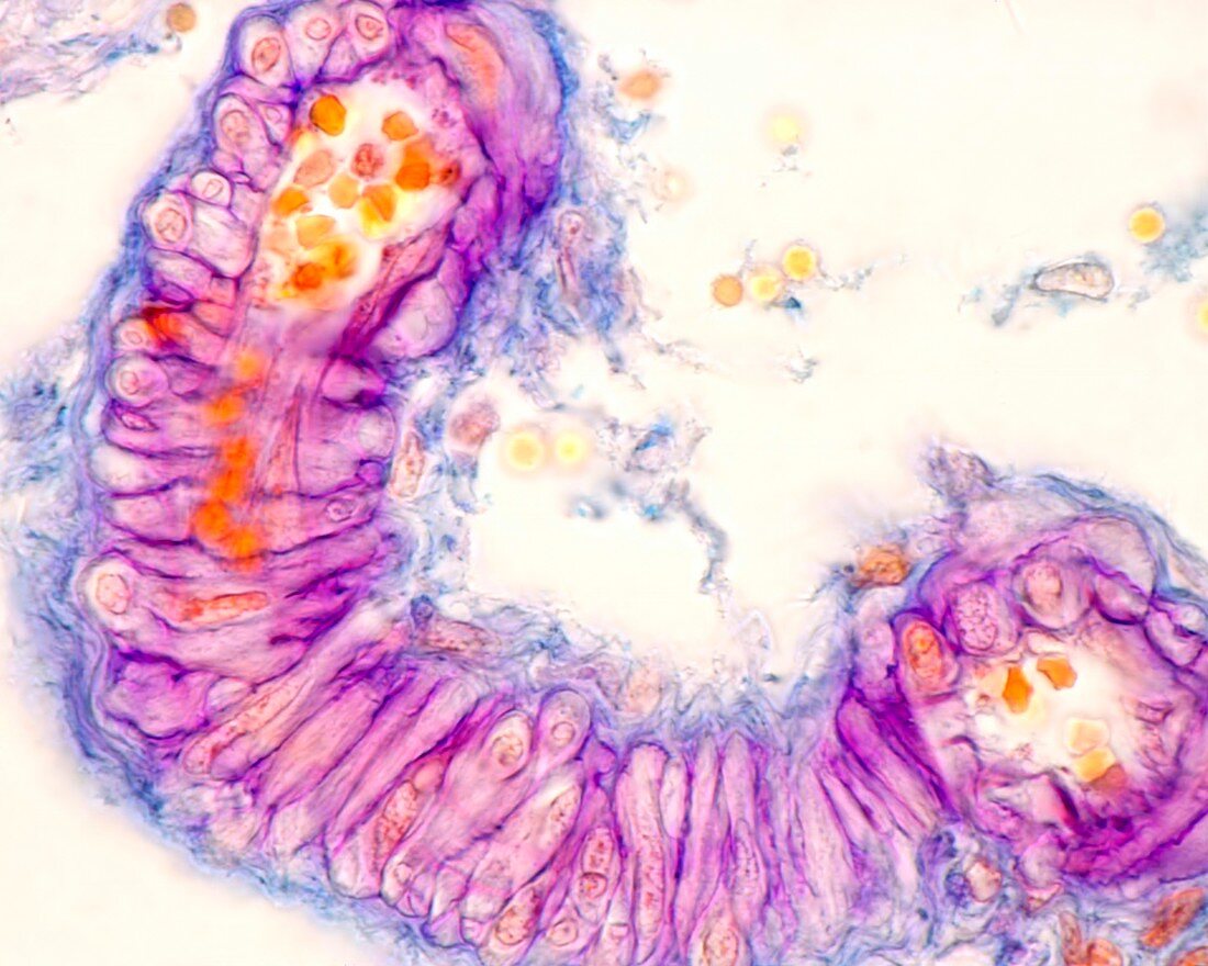
| Light micrograph of a longitudinal section of an arteriole stained with the Gabe's aldehyde-fucsin method. This method stains the basement membranes surrounding the smooth muscle cells of the tunica media. Due to the incurved path of the vessel, these cells appear sectioned longitudinally in the central zona and in cross section at the ends. Magnification: x900 when printed at 10 centimetres across. | |
| Lizenzart: | Lizenzpflichtig |
| Credit: | Science Photo Library / JOSE CALVO |
| Bildgröße: | 4674 px × 3739 px |
| Modell-Rechte: | nicht erforderlich |
| Eigentums-Rechte: | nicht erforderlich |
| Restrictions: | - |
Preise für dieses Bild ab 15 €
Universitäten & Organisationen
(Informationsmaterial Digital, Informationsmaterial Print, Lehrmaterial Digital etc.)
ab 15 €
Redaktionell
(Bücher, Bücher: Sach- und Fachliteratur, Digitale Medien (redaktionell) etc.)
ab 30 €
Werbung
(Anzeigen, Aussenwerbung, Digitale Medien, Fernsehwerbung, Karten, Werbemittel, Zeitschriften etc.)
ab 55 €
Handelsprodukte
(bedruckte Textilie, Kalender, Postkarte, Grußkarte, Verpackung etc.)
ab 75 €
Pauschalpreise
Rechtepakete für die unbeschränkte Bildnutzung in Print oder Online
ab 495 €
Keywords
- Adventitia,
- Anatomie,
- anatomisch,
- Arterie,
- arteriell,
- Arteriole,
- Ballaststoff,
- befleckt,
- Bindegewebe,
- Biologie,
- biologisch,
- Blut,
- Blutfluss,
- Blutgefäß,
- Blutgefäße,
- Blutzelle,
- Elastin,
- elastische Fasern,
- Fasern,
- Gefäß,
- Gefäße,
- gesund,
- Gesundheit,
- Gesundheitswesen,
- Glatt,
- Histologie,
- histologisch,
- Kreislauf,
- Lichtmikroskop,
- lichtmikroskopische Aufnahme,
- Medien,
- Mensch,
- menschlicher Körper,
- menschliches Gewebe,
- Muskulös,
- normal,
- rote Blutkörperchen,
- Schicht,
- Schichten,
- Sektion,
- Tunica intima,
- vaskulär,
- Vene