Prostate cancer, abdominal CT scan
Bildnummer 12303351
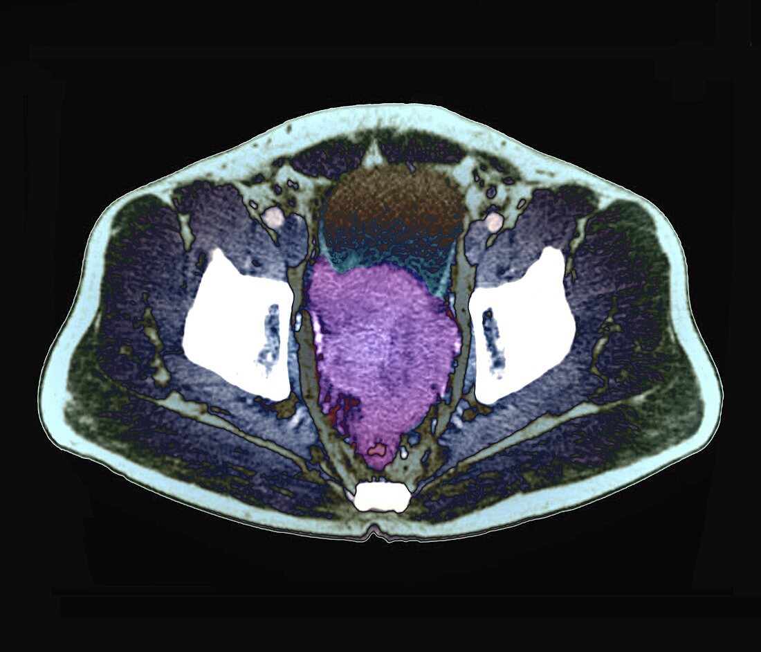
| Prostate cancer. Coloured axial computed tomography (CT) scan through the abdomen of a 88-year-old man with prostate cancer. The scan is centred on the bladder, with the spine (white, lower centre) and hip bones (white, left and right) also visible. At centre is a large malignant tumour (purple) that has formed in the prostate gland and spread to the rectum and the bladder wall. | |
| Lizenzart: | Lizenzpflichtig |
| Credit: | Science Photo Library / Zephyr |
| Bildgröße: | 4518 px × 3868 px |
| Modell-Rechte: | nicht erforderlich |
| Eigentums-Rechte: | nicht erforderlich |
| Restrictions: | - |
Preise für dieses Bild ab 15 €
Universitäten & Organisationen
(Informationsmaterial Digital, Informationsmaterial Print, Lehrmaterial Digital etc.)
ab 15 €
Redaktionell
(Bücher, Bücher: Sach- und Fachliteratur, Digitale Medien (redaktionell) etc.)
ab 30 €
Werbung
(Anzeigen, Aussenwerbung, Digitale Medien, Fernsehwerbung, Karten, Werbemittel, Zeitschriften etc.)
ab 55 €
Handelsprodukte
(bedruckte Textilie, Kalender, Postkarte, Grußkarte, Verpackung etc.)
ab 75 €
Pauschalpreise
Rechtepakete für die unbeschränkte Bildnutzung in Print oder Online
ab 495 €
Keywords
- Abdomen,
- abnormal,
- achtziger Jahre,
- Alt,
- älter,
- ausgeschnitten,
- Ausschnitte,
- axial,
- Bauch,
- Blase,
- Computertomographie,
- CT-Scan,
- Drüse,
- Enddarm,
- Erwachsene,
- farbig,
- geduldig,
- gefärbt,
- Gewebe,
- Kondition,
- Krankheit,
- Krebs,
- krebsartig,
- maligne,
- Malignom,
- Mann,
- Männlich,
- Medizin,
- medizinisch,
- menschlicher Körper,
- Niemand,
- Onkologie,
- Organ,
- primär,
- Prostata,
- Prostatakrebs,
- Scanner,
- schwarzer Hintergrund,
- Sektion,
- sektioniert,
- sekundär,
- Störung,
- Tumor,
- ungesund,
- Verbreitung,
- Wachstum