Human brain, coronal MRI scan
Bildnummer 12303275
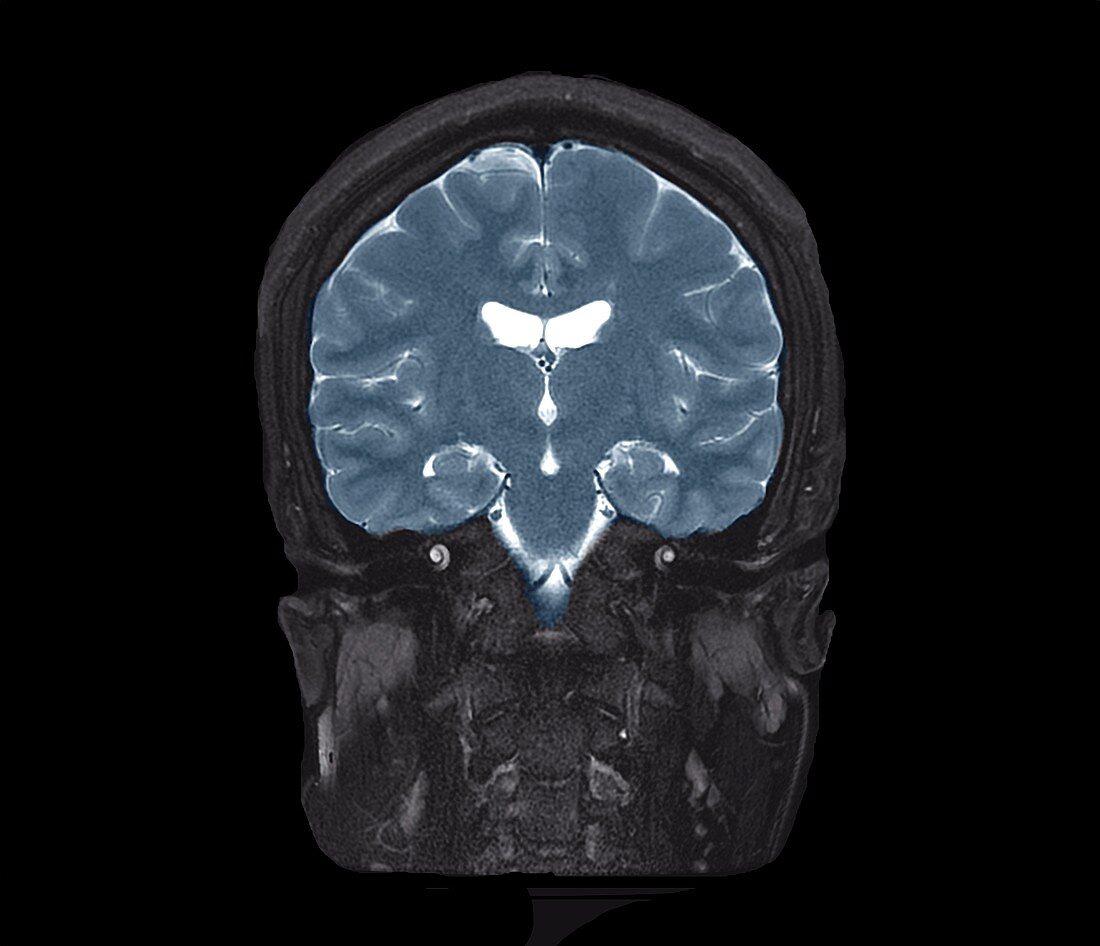
| Human brain. Coloured coronal magnetic resonance imaging (MRI) scan through the brain of a 35-year-old man. This is a T2-weighted scan, with injection of gadolinium contrast medium. The scan has passed vertically through the head, as seen from the front, at the level of the ears. The scan shows the normal brain structures, including parts of the lateral ventricles (white, centre) and the folds of the cerebrum. Below the brain, on either side of the head, are the structures of the inner ear. Parts of the cervical spine are also seen in the neck. | |
| Lizenzart: | Lizenzpflichtig |
| Credit: | Science Photo Library / Zephyr |
| Bildgröße: | 4508 px × 3876 px |
| Modell-Rechte: | nicht erforderlich |
| Eigentums-Rechte: | nicht erforderlich |
| Restrictions: | - |
Preise für dieses Bild ab 15 €
Universitäten & Organisationen
(Informationsmaterial Digital, Informationsmaterial Print, Lehrmaterial Digital etc.)
ab 15 €
Redaktionell
(Bücher, Bücher: Sach- und Fachliteratur, Digitale Medien (redaktionell) etc.)
ab 30 €
Werbung
(Anzeigen, Aussenwerbung, Digitale Medien, Fernsehwerbung, Karten, Werbemittel, Zeitschriften etc.)
ab 55 €
Handelsprodukte
(bedruckte Textilie, Kalender, Postkarte, Grußkarte, Verpackung etc.)
ab 75 €
Pauschalpreise
Rechtepakete für die unbeschränkte Bildnutzung in Print oder Online
ab 495 €
Keywords
- 30er Jahre,
- Anatomie,
- anatomisch,
- ausgeschnitten,
- Ausschnitte,
- Biologie,
- biologisch,
- dreißiger Jahre,
- Erwachsene,
- farbig,
- gefärbt,
- Gehirn,
- gesund,
- Hals,
- Halswirbelsäule,
- Innenohr,
- Knochen,
- Kopf,
- Magnetresonanztomografie,
- Mann,
- Männlich,
- menschlicher Körper,
- MRT-Untersuchung,
- Niemand,
- normal,
- Ohr,
- Ohren,
- Organ,
- Scanner,
- Schädel,
- schwarzer Hintergrund,
- Sektion,
- sektioniert,
- Stirnbein,
- Ventrikel,
- Wirbelsäule,
- Wirbelsäulen-