Knee ligaments, illustration
Bildnummer 12287353
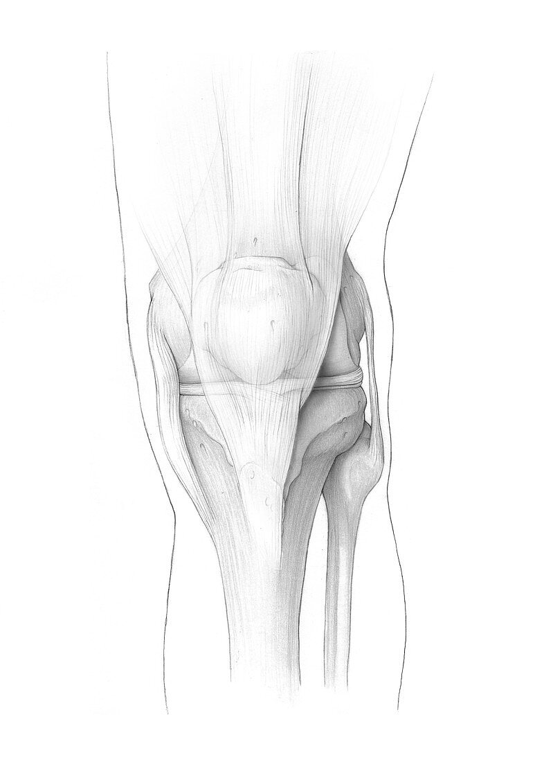
| Illustration showing ligament anatomy of a straight human knee. Ligaments are fibrous bands of connective tissue that connect bones. At the centre of the knee is the patella (kneecap). The patella tendon attaches the bottom of the patella to the tibia (shin bone). On the inside of the knee (left) is the medial collateral ligament (MCL), which connects the femur (thigh bone) and tibia and limits sideways movement. The fibular collateral ligament (FCL) performs the same function on the outside of the knee, by connecting the femur and fibula (calf bone, bottom right). In the centre of the knee joint are the anterior cruciate ligament (ACL), and posterior cruciate ligament (PCL), which connect the femur and tibia. They prevent forwards and backwards movements (respectively) of the bones in relation to one another. Running horizontally across the joint are the menisci (medial meniscus at left and lateral meniscus at right), which act as shock absorbers for the knee. | |
| Lizenzart: | Lizenzpflichtig |
| Credit: | Science Photo Library / VERONICA FALCONIERI HAYS |
| Bildgröße: | 3729 px × 5220 px |
| Modell-Rechte: | nicht erforderlich |
| Eigentums-Rechte: | nicht erforderlich |
| Restrictions: | - |
Preise für dieses Bild ab 15 €
Universitäten & Organisationen
(Informationsmaterial Digital, Informationsmaterial Print, Lehrmaterial Digital etc.)
ab 15 €
Redaktionell
(Bücher, Bücher: Sach- und Fachliteratur, Digitale Medien (redaktionell) etc.)
ab 30 €
Werbung
(Anzeigen, Aussenwerbung, Digitale Medien, Fernsehwerbung, Karten, Werbemittel, Zeitschriften etc.)
ab 55 €
Handelsprodukte
(bedruckte Textilie, Kalender, Postkarte, Grußkarte, Verpackung etc.)
ab 75 €
Pauschalpreise
Rechtepakete für die unbeschränkte Bildnutzung in Print oder Online
ab 495 €