Cilia-based defects cancer research
Bildnummer 12248850
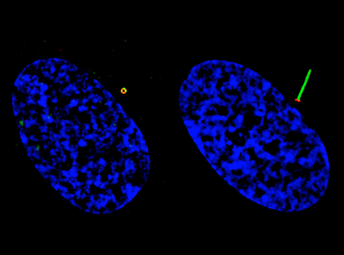
| Cilia-based defects cancer research. Structured illumination microscopy (SIM) imaging of two human retinal pigment epithelium (RPE) cells, showing ciliogenesis progression (the formation of cilia). Defects in ciliary formation and function are linked to several human diseases, including cancer. Ciliogenesis starts from docking pre-ciliary vesicles to the distal appendage of the mother centriole. Those docked vesicles fuse to form a larger ciliary vesicle and then extend to the ciliary membrane. At left, GFP-Smoothened (green) vesicles are docked at the mother centriole/basal body distal appendage (marked by CEP164 in red). At right, ciliary GFP-Smoothened (green) is localized on the ciliary membrane. The cell nuclei are shown in blue. | |
| Lizenzart: | Lizenzpflichtig |
| Credit: | Science Photo Library / NATIONAL CANCER INSTITUTE / NCI Center for Cancer Research |
| Bildgröße: | 3476 px × 2575 px |
| Modell-Rechte: | nicht erforderlich |
| Eigentums-Rechte: | nicht erforderlich |
| Restrictions: |
|
Preise für dieses Bild ab 15 €
Universitäten & Organisationen
(Informationsmaterial Digital, Informationsmaterial Print, Lehrmaterial Digital etc.)
ab 15 €
Redaktionell
(Bücher, Bücher: Sach- und Fachliteratur, Digitale Medien (redaktionell) etc.)
ab 30 €
Werbung
(Anzeigen, Aussenwerbung, Digitale Medien, Fernsehwerbung, Karten, Werbemittel, Zeitschriften etc.)
ab 55 €
Handelsprodukte
(bedruckte Textilie, Kalender, Postkarte, Grußkarte, Verpackung etc.)
ab 75 €
Pauschalpreise
Rechtepakete für die unbeschränkte Bildnutzung in Print oder Online
ab 495 €
Keywords
- abnormal,
- Duo,
- Forschung,
- Gewebe,
- GFP,
- Grün fluoreszierendes Protein,
- Kondition,
- Krankheit,
- Krebs,
- krebsartig,
- Krebszelle,
- Lichtmikroskop,
- lichtmikroskopische Aufnahme,
- maligne,
- Malignom,
- Medizin,
- medizinisch,
- Niemand,
- Onkologie,
- Paar,
- Proteine,
- retinales Pigmentepithel,
- rpe,
- schwarzer Hintergrund,
- Störung,
- Tumor,
- ungesund,
- Wimpern,
- Zelle,
- Zellen,
- zellular,
- Zwei