HeLa cells with Adenovirus
Bildnummer 12066061
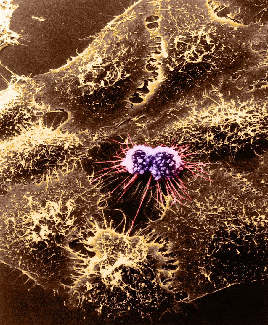
| Coloured scanning electron micrograph (SEM) of cultured HeLa cells originally derived many years ago from a womans cancerous cervical tissue. A light micrograph (x130) of the same cells (inset) reveals rounded double cells in the center in the process of dividing. This HeLa cell (named after patient Henrietta Lacks) has been infected with adenovirus (a DNA virus). After 4-1/2 hours the HeLa cells surface becomes rough. The multiple surface blebs on this cell are characteristic for a certain stage of cell division that both normal and cancer cells go through. Research with the SEM has established the extraordinarily responsive nature of cell surface form. This instrument records,in pictures,specific cell reactions to various changes in the cells environment and maps the distribution of surface binding sites for biologically important molecules such as hormone,antigens,and pharmacologic agents | |
| Lizenzart: | Lizenzpflichtig |
| Credit: | Science Photo Library / Science Source |
| Bildgröße: | 2468 px × 3000 px |
| Modell-Rechte: | nicht erforderlich |
| Eigentums-Rechte: | nicht erforderlich |
| Restrictions: |
|
Preise für dieses Bild ab 15 €
Universitäten & Organisationen
(Informationsmaterial Digital, Informationsmaterial Print, Lehrmaterial Digital etc.)
ab 15 €
Redaktionell
(Bücher, Bücher: Sach- und Fachliteratur, Digitale Medien (redaktionell) etc.)
ab 30 €
Werbung
(Anzeigen, Aussenwerbung, Digitale Medien, Fernsehwerbung, Karten, Werbemittel, Zeitschriften etc.)
ab 55 €
Handelsprodukte
(bedruckte Textilie, Kalender, Postkarte, Grußkarte, Verpackung etc.)
ab 75 €
Pauschalpreise
Rechtepakete für die unbeschränkte Bildnutzung in Print oder Online
ab 495 €