Colour Enhanced Severe Alzheimer's Diseas
Bildnummer 12031010
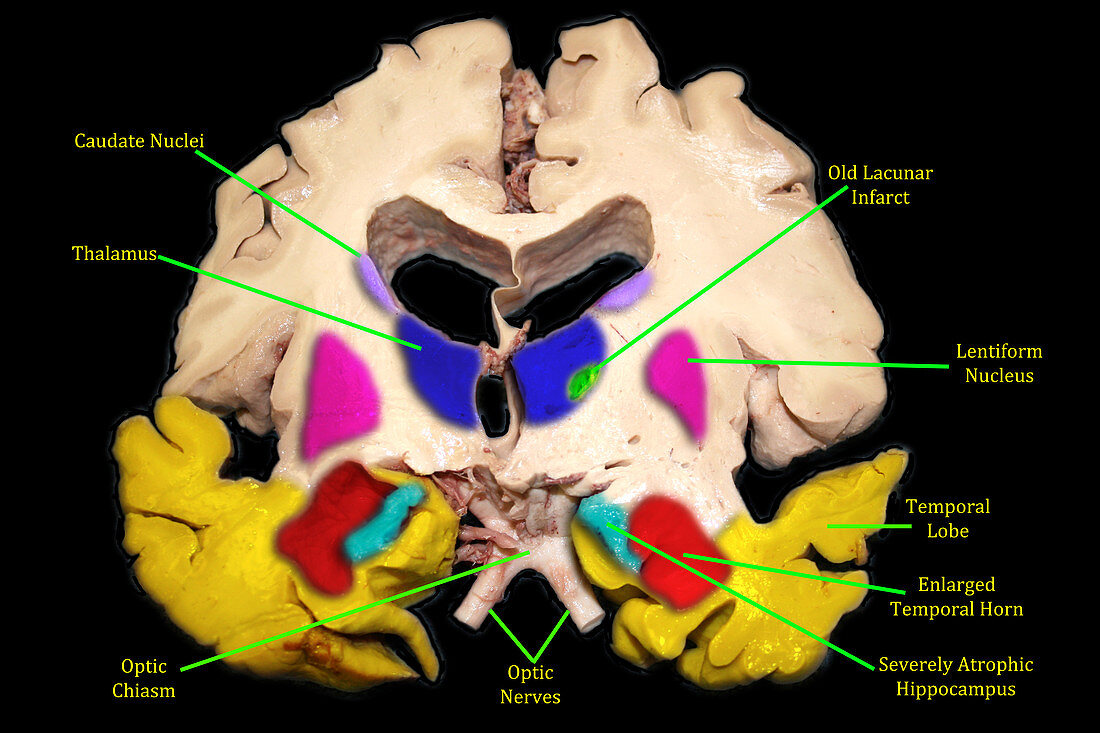
| This colour enhanced and labelled coronal (frontal view) gross anatomic brain specimen demonstrates evidence of severe,endstage Alzheimer's disease with marked atrophy of the hippocampal formations (aqua) (which are very important for memory) with associated enlargement of the temporal horns (red) of the lateral ventricles. There is also prominent generalized atrophy of the remaining portions of the temporal lobes (yellow) with enlargement of the sylvian fissures. There is also atrophy with enlargement of the third ventricle and bodies of the lateral ventricles. The lentiform nucleus of the basal ganglia is pink. The thalamus is dark blue with a small lacunar infarct in the left (on viewers right) thalamus. The caudate nuclei of the basal ganglia are | |
| Lizenzart: | Lizenzpflichtig |
| Credit: | Science Photo Library / Living Art Enterprises |
| Bildgröße: | 5400 px × 3600 px |
| Modell-Rechte: | nicht erforderlich |
| Eigentums-Rechte: | nicht erforderlich |
| Restrictions: |
|
Preise für dieses Bild ab 15 €
Universitäten & Organisationen
(Informationsmaterial Digital, Informationsmaterial Print, Lehrmaterial Digital etc.)
ab 15 €
Redaktionell
(Bücher, Bücher: Sach- und Fachliteratur, Digitale Medien (redaktionell) etc.)
ab 30 €
Werbung
(Anzeigen, Aussenwerbung, Digitale Medien, Fernsehwerbung, Karten, Werbemittel, Zeitschriften etc.)
ab 55 €
Handelsprodukte
(bedruckte Textilie, Kalender, Postkarte, Grußkarte, Verpackung etc.)
ab 75 €
Pauschalpreise
Rechtepakete für die unbeschränkte Bildnutzung in Print oder Online
ab 495 €