Abdominal Aorta
Bildnummer 12006783
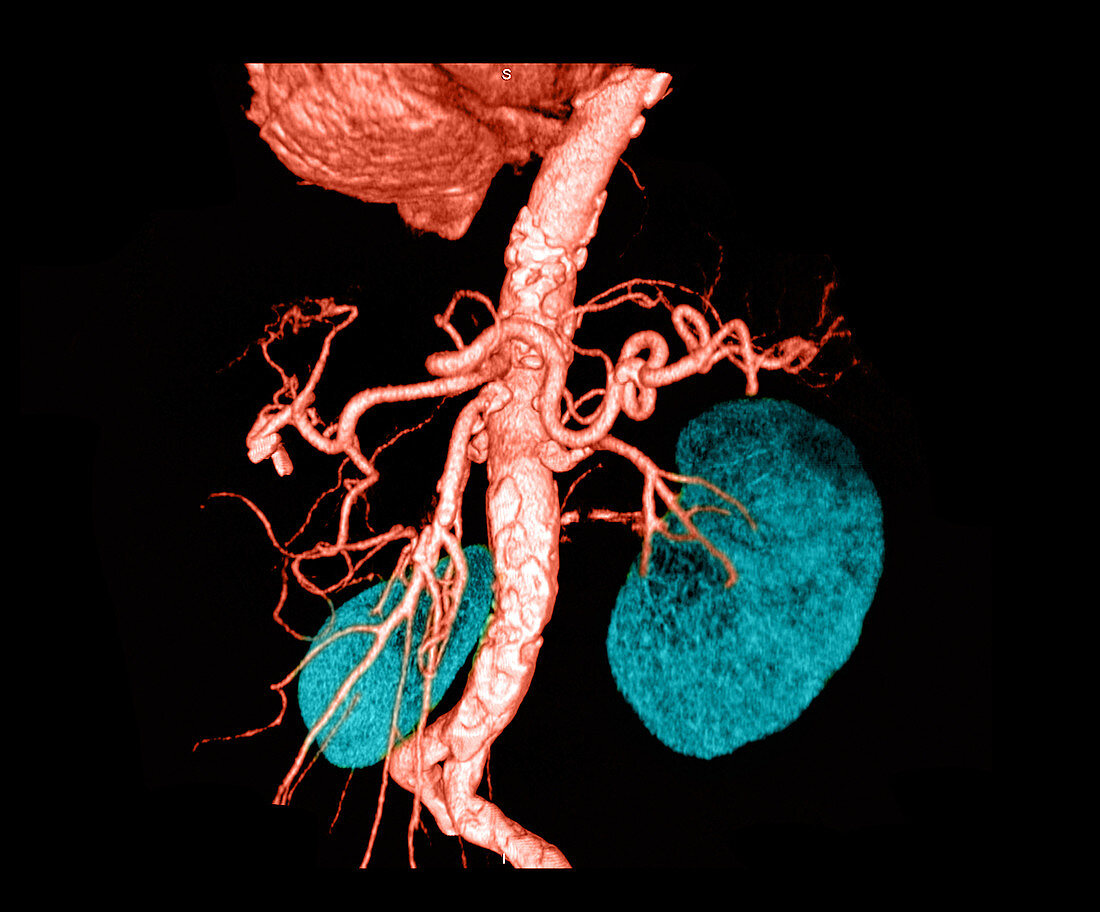
| This frontal view from a 3D reconstruction of the abdominal aorta was obtained utilizing data from a CT angiogram. This image shows the bottom of the heart at the top of the image. The celiac axis and the superior mesenteric artery branches of the abdominal aorta are seen in addition to the renal arteries and both kidneys (blue). The irregularity along the walls of the vessels are a result of atherosclerotic plaque | |
| Lizenzart: | Lizenzpflichtig |
| Credit: | Science Photo Library / Medical Body Scans |
| Bildgröße: | 3617 px × 3000 px |
| Modell-Rechte: | nicht erforderlich |
| Eigentums-Rechte: | nicht erforderlich |
| Restrictions: |
|
Preise für dieses Bild ab 15 €
Universitäten & Organisationen
(Informationsmaterial Digital, Informationsmaterial Print, Lehrmaterial Digital etc.)
ab 15 €
Redaktionell
(Bücher, Bücher: Sach- und Fachliteratur, Digitale Medien (redaktionell) etc.)
ab 30 €
Werbung
(Anzeigen, Aussenwerbung, Digitale Medien, Fernsehwerbung, Karten, Werbemittel, Zeitschriften etc.)
ab 55 €
Handelsprodukte
(bedruckte Textilie, Kalender, Postkarte, Grußkarte, Verpackung etc.)
ab 75 €
Pauschalpreise
Rechtepakete für die unbeschränkte Bildnutzung in Print oder Online
ab 495 €
Keywords
- 3 dimensional,
- 3D,
- Anatomie,
- Angiogramm,
- Aorta,
- Blutgefäß,
- Computertomographie,
- CTA,
- Diagnose,
- diagnostische Bildgebung,
- diagnostizieren,
- Dreidimensional,
- eingefärbt,
- Farbe,
- farbverstärkt,
- Gefäßsystem,
- gesund,
- Kolorieren,
- Kreislauf,
- Medizin,
- medizinisch,
- medizinische Bildgebung,
- medizinischer Scan,
- medizinisches Bild,
- Niere,
- normal,
- Scan,
- Wissenschaft