Gem anemone,light micrograph
Bildnummer 11906360
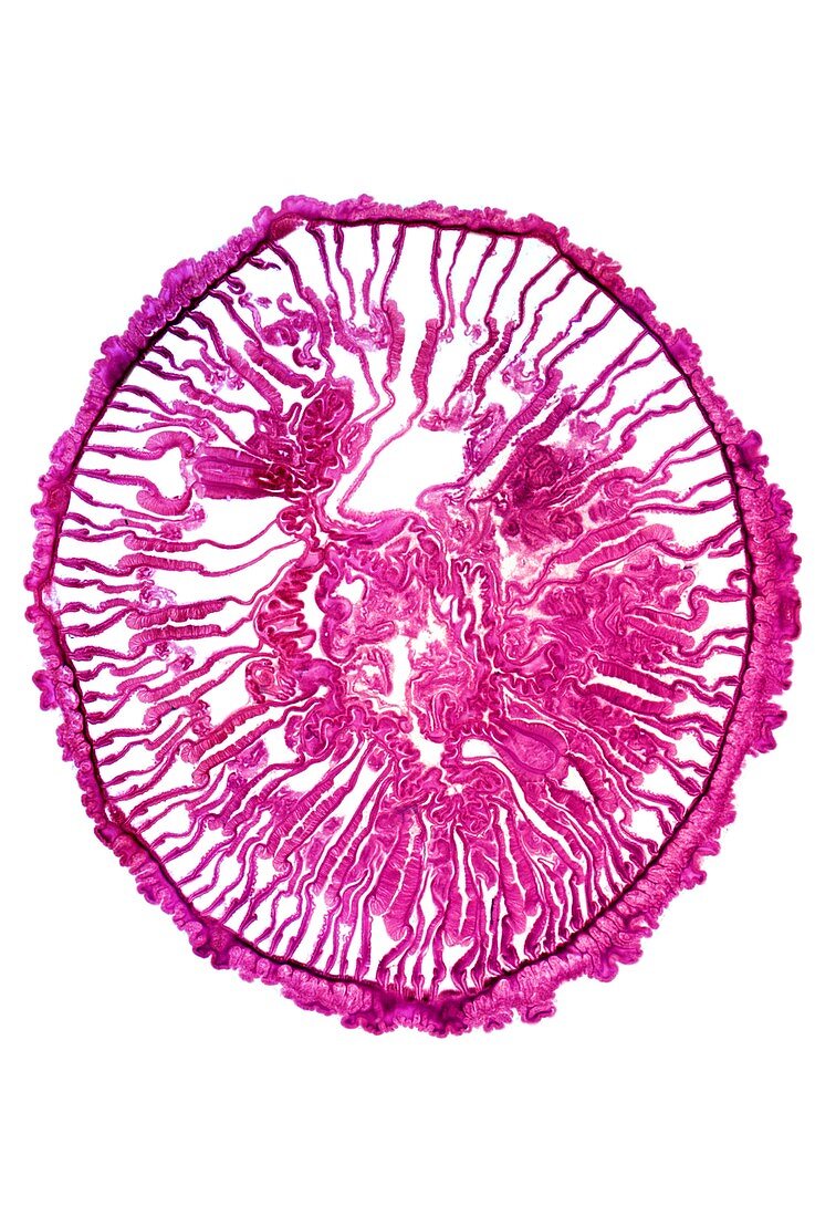
| Gem anemone. Light micrograph of a transverse section through the body of a gem anemone (Bunodactis verrucosa). This section has passed through the pharynx (stomodaeum) region,showing the outer and inner epidermal walls,which are joined by septae (mesenteries,lines radiating inwards) which divide the anemone's body into compartments. Attached to the mesenteries are longitudinal bands of muscle (feathery areas). These retractor muscles contract to shorten the body and invert the tentacles (polyps) forcing water in and out of the body cavities. Magnification: x14 when printed at 10 centimetres high | |
| Lizenzart: | Lizenzpflichtig |
| Credit: | Science Photo Library / Wheeler, Dr. Keith |
| Bildgröße: | 3761 px × 5616 px |
| Modell-Rechte: | nicht erforderlich |
| Eigentums-Rechte: | nicht erforderlich |
| Restrictions: | - |
Preise für dieses Bild ab 15 €
Universitäten & Organisationen
(Informationsmaterial Digital, Informationsmaterial Print, Lehrmaterial Digital etc.)
ab 15 €
Redaktionell
(Bücher, Bücher: Sach- und Fachliteratur, Digitale Medien (redaktionell) etc.)
ab 30 €
Werbung
(Anzeigen, Aussenwerbung, Digitale Medien, Fernsehwerbung, Karten, Werbemittel, Zeitschriften etc.)
ab 55 €
Handelsprodukte
(bedruckte Textilie, Kalender, Postkarte, Grußkarte, Verpackung etc.)
ab 75 €
Pauschalpreise
Rechtepakete für die unbeschränkte Bildnutzung in Print oder Online
ab 495 €
Keywords
- Abteil,
- Anatomie,
- anatomisch,
- Anemone,
- ausgeschnitten,
- Ausschnitte,
- Biologie,
- biologisch,
- diagonal,
- einer,
- epidermal,
- Epidermis,
- Fauna,
- Histologie,
- histologisch,
- Hohltier,
- Kreis,
- kreisförmig,
- Lichtmikroskop,
- lichtmikroskopische Aufnahme,
- Mauer,
- Meeresbiologie,
- Natur,
- Polyp,
- Polypen,
- Querschnitt,
- rund,
- Sektion,
- sektioniert,
- Single,
- Tentakel,
- Tentakeln,
- Tier,
- Tierwelt,
- Wände,
- wirbellos,
- Wirbellose,
- Zoologie,
- zoologisch