First whole body X-ray,1897
Bildnummer 11905359
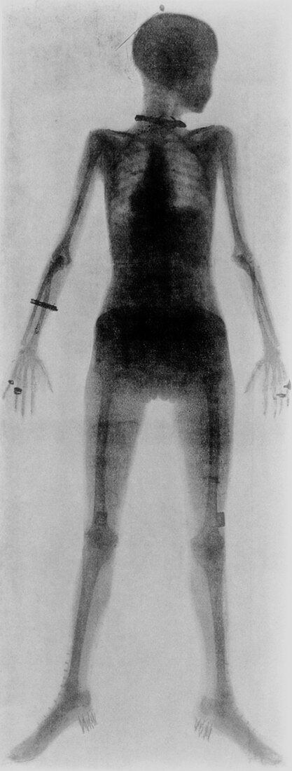
| First whole body X-ray (1907). The first whole body radiograph ever taken of a living person,a woman in 1897. The X-ray was made from a single exposure by William Morton of New York. Standing with her head in profile,her skeleton,heart and liver are seen. Her jewellery is highly visible: hatpin,necklace,bracelet,rings. On her feet she is wearing high button boots with nailed-on heels,and around her hips and abdomen a whalebone corset. To make this X-ray,Morton included a 12 inch induction coil with the current supplied from a New York street mains connection. A Crookes' tube was positioned 54 inches from the X-ray plate and the time taken was about 30 minutes | |
| Lizenzart: | Lizenzpflichtig |
| Credit: | Science Photo Library |
| Bildgröße: | 1863 px × 4919 px |
| Modell-Rechte: | nicht erforderlich |
| Eigentums-Rechte: | nicht erforderlich |
| Restrictions: | - |
Preise für dieses Bild ab 15 €
Universitäten & Organisationen
(Informationsmaterial Digital, Informationsmaterial Print, Lehrmaterial Digital etc.)
ab 15 €
Redaktionell
(Bücher, Bücher: Sach- und Fachliteratur, Digitale Medien (redaktionell) etc.)
ab 30 €
Werbung
(Anzeigen, Aussenwerbung, Digitale Medien, Fernsehwerbung, Karten, Werbemittel, Zeitschriften etc.)
ab 55 €
Handelsprodukte
(bedruckte Textilie, Kalender, Postkarte, Grußkarte, Verpackung etc.)
ab 75 €
Pauschalpreise
Rechtepakete für die unbeschränkte Bildnutzung in Print oder Online
ab 495 €