Mitosis cell division
Bildnummer 11874821
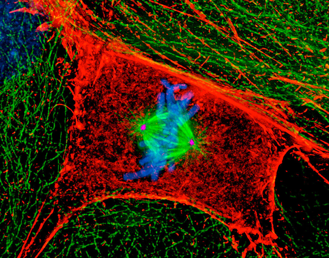
| Mitosis (image 3 of 6). Digital three-dimensional immunofluorescent light micrograph of a section through a mammalian cell during the metaphase stage of mitotic cell division. The chromosomes (blue) have lined up along the centre of the cell. The actin microfilaments (red) and tubulin microtubules (green) of the cytoskeleton maintain the cell's structure. The two centrosomes (pink dots,centre) organize the microtubules that move the chromosomes around the cell during mitosis. Mitosis produces two identical daughter cells. This is a rat kangaroo kidney epithelial cell. Magnification: x500 at 6x7cm size. For a sequence of mitosis see images P673/052-057 | |
| Lizenzart: | Lizenzpflichtig |
| Credit: | Science Photo Library / Khodjakov, Dr. Alexey |
| Bildgröße: | 3500 px × 2742 px |
| Modell-Rechte: | nicht erforderlich |
| Eigentums-Rechte: | nicht erforderlich |
| Restrictions: | - |
Preise für dieses Bild ab 15 €
Universitäten & Organisationen
(Informationsmaterial Digital, Informationsmaterial Print, Lehrmaterial Digital etc.)
ab 15 €
Redaktionell
(Bücher, Bücher: Sach- und Fachliteratur, Digitale Medien (redaktionell) etc.)
ab 30 €
Werbung
(Anzeigen, Aussenwerbung, Digitale Medien, Fernsehwerbung, Karten, Werbemittel, Zeitschriften etc.)
ab 55 €
Handelsprodukte
(bedruckte Textilie, Kalender, Postkarte, Grußkarte, Verpackung etc.)
ab 75 €
Pauschalpreise
Rechtepakete für die unbeschränkte Bildnutzung in Print oder Online
ab 495 €