Mitosis cell division
Bildnummer 11874819
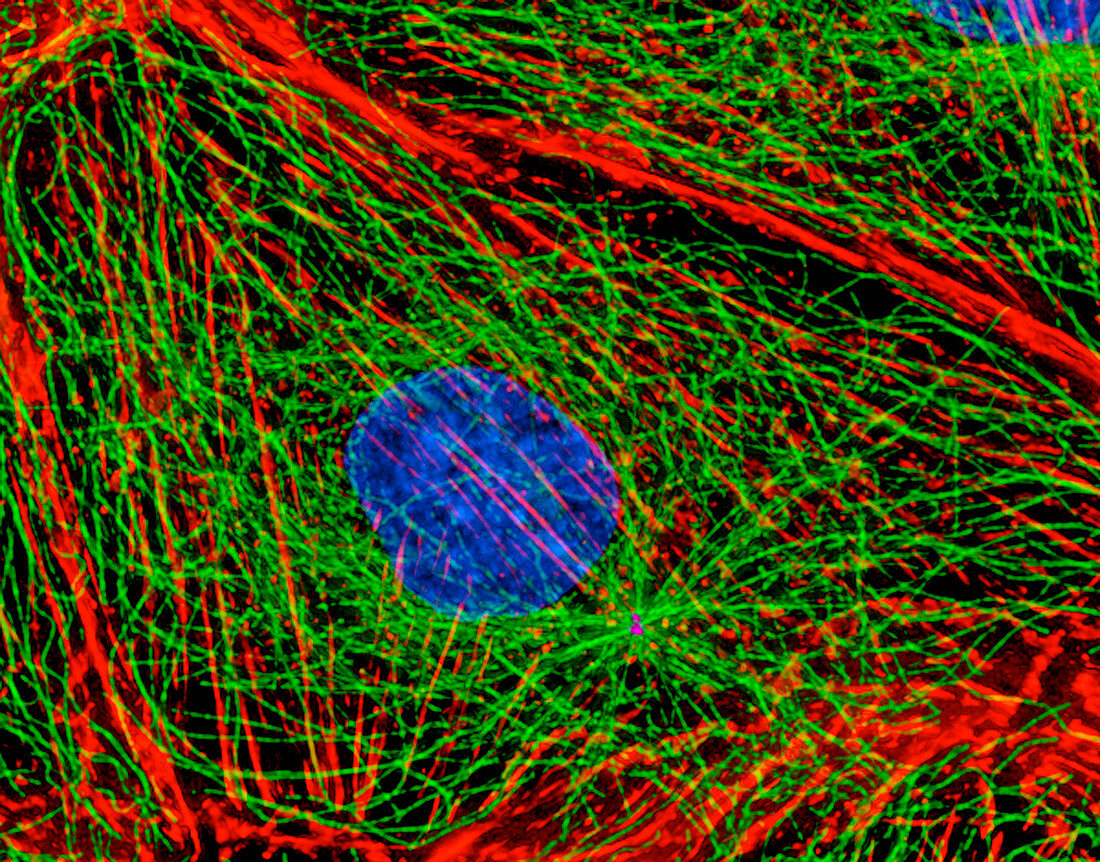
| Mitosis (image 1 of 6). Digital three-dimensional immunofluorescent light micrograph of a section through a mammalian cell during interphase,the stage between mitotic cell divisions. The chromosomes (blue) replicate during this stage. The actin microfilaments (red) and tubulin microtubules (green) of the cytoskeleton maintain the structure and organisation of the cell. Mitosis results in the formation of two identical daughter cells. Antibodies have been used to attach fluorescent dyes to specific cell tissues. This is a kidney epithelial cell from a rat kangaroo. Magnification: x500 at 6x7cm size. For a sequence of mitosis see images P673/052-057 | |
| Lizenzart: | Lizenzpflichtig |
| Credit: | Science Photo Library / Khodjakov, Dr. Alexey |
| Bildgröße: | 3500 px × 2742 px |
| Modell-Rechte: | nicht erforderlich |
| Eigentums-Rechte: | nicht erforderlich |
| Restrictions: | - |
Preise für dieses Bild ab 15 €
Universitäten & Organisationen
(Informationsmaterial Digital, Informationsmaterial Print, Lehrmaterial Digital etc.)
ab 15 €
Redaktionell
(Bücher, Bücher: Sach- und Fachliteratur, Digitale Medien (redaktionell) etc.)
ab 30 €
Werbung
(Anzeigen, Aussenwerbung, Digitale Medien, Fernsehwerbung, Karten, Werbemittel, Zeitschriften etc.)
ab 55 €
Handelsprodukte
(bedruckte Textilie, Kalender, Postkarte, Grußkarte, Verpackung etc.)
ab 75 €
Pauschalpreise
Rechtepakete für die unbeschränkte Bildnutzung in Print oder Online
ab 495 €