Artwork showing structure of small intestine villi
Bildnummer 11872669
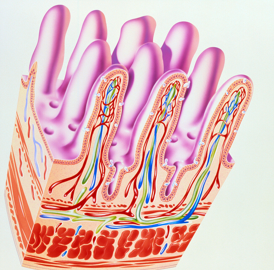
| Small intesine villi. Illustration of the finger- like villi that line the inside wall of the human small intestine. Three villi are shown in section. Inside them is a network of blood vessels (red and blue) that absorbs the products of protein and carbohydrate digestion. The green vessels are branches of the lymphatic system called lacteals. These absorb the products of fat digestion. A layer of epithelial cells covers the villi. Among these are goblet cells (white),which produce a lubricating mucus. The cavities between villi are the crypts of Lieberkuhn. These contain glands that secrete digestive enzymes. The layers of muscle (bottom) squeeze food through the gut | |
| Lizenzart: | Lizenzpflichtig |
| Credit: | Science Photo Library / Bavosi, John |
| Bildgröße: | 3087 px × 3046 px |
| Modell-Rechte: | nicht erforderlich |
| Eigentums-Rechte: | nicht erforderlich |
| Restrictions: | - |
Preise für dieses Bild ab 15 €
Universitäten & Organisationen
(Informationsmaterial Digital, Informationsmaterial Print, Lehrmaterial Digital etc.)
ab 15 €
Redaktionell
(Bücher, Bücher: Sach- und Fachliteratur, Digitale Medien (redaktionell) etc.)
ab 30 €
Werbung
(Anzeigen, Aussenwerbung, Digitale Medien, Fernsehwerbung, Karten, Werbemittel, Zeitschriften etc.)
ab 55 €
Handelsprodukte
(bedruckte Textilie, Kalender, Postkarte, Grußkarte, Verpackung etc.)
ab 75 €
Pauschalpreise
Rechtepakete für die unbeschränkte Bildnutzung in Print oder Online
ab 495 €