Functional map of the brain's motor cortex areas
Bildnummer 11870923
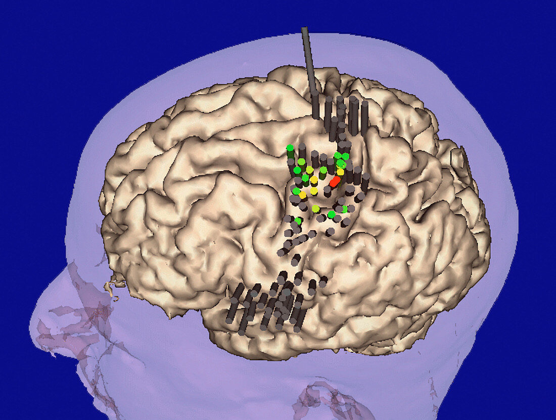
| Motor cortex brain areas. 3-D magnetic resonance imaging (MRI) scan of the brain mapped to reveal areas of the cerebral motor cortex,which controls voluntary muscle activity. This mapping is used in virtual reality assisted surgery. Electric currents are first applied to parts of the motor cortex to trigger muscle responses in the body and create a map. The map is shown by colour-coded cylinders which give intensity of response: red (strong),yellow (medium),green (weak) and grey (no response). Surgery,such as tumour removal,can then be done without risking damage to the motor cortex | |
| Lizenzart: | Lizenzpflichtig |
| Credit: | Science Photo Library / BRIGHAM & WOMEN'S HOSPITAL / SURGICAL PLANNING LAB / MIT AI LAB |
| Bildgröße: | 3543 px × 2672 px |
| Modell-Rechte: | nicht erforderlich |
| Eigentums-Rechte: | nicht erforderlich |
| Restrictions: | - |
Preise für dieses Bild ab 15 €
Universitäten & Organisationen
(Informationsmaterial Digital, Informationsmaterial Print, Lehrmaterial Digital etc.)
ab 15 €
Redaktionell
(Bücher, Bücher: Sach- und Fachliteratur, Digitale Medien (redaktionell) etc.)
ab 30 €
Werbung
(Anzeigen, Aussenwerbung, Digitale Medien, Fernsehwerbung, Karten, Werbemittel, Zeitschriften etc.)
ab 55 €
Handelsprodukte
(bedruckte Textilie, Kalender, Postkarte, Grußkarte, Verpackung etc.)
ab 75 €
Pauschalpreise
Rechtepakete für die unbeschränkte Bildnutzung in Print oder Online
ab 495 €