Illustration showing the nerves of the human heart
Bildnummer 11868970
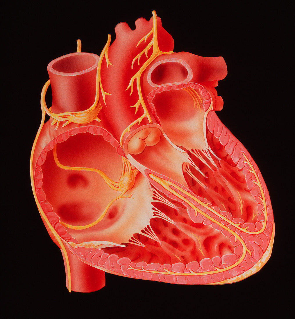
| Illustration of a section through the human heart,showing (in yellow) the nerves that govern the pumping cycle. Blood flows from right and left ventricles (bottom left & right respectively on image). The electrical impulse that provokes ventricular contraction begins in the Sinoatrial (SA) node,the heart's pacemaker (yellow bundle at top left),spreading to both left & right atria (top) and down to the atrioventricular (AV) node (located between right atrium & ventricle). The AV node delays passage of the impulse to allow time for the ventricles to fill before effecting ventricular contraction via branches of the bundle of HIS (yellow fibres between ventricles) | |
| Lizenzart: | Lizenzpflichtig |
| Credit: | Science Photo Library / Gifford, David |
| Bildgröße: | 3622 px × 3925 px |
| Modell-Rechte: | nicht erforderlich |
| Eigentums-Rechte: | nicht erforderlich |
| Restrictions: | - |
Preise für dieses Bild ab 15 €
Universitäten & Organisationen
(Informationsmaterial Digital, Informationsmaterial Print, Lehrmaterial Digital etc.)
ab 15 €
Redaktionell
(Bücher, Bücher: Sach- und Fachliteratur, Digitale Medien (redaktionell) etc.)
ab 30 €
Werbung
(Anzeigen, Aussenwerbung, Digitale Medien, Fernsehwerbung, Karten, Werbemittel, Zeitschriften etc.)
ab 55 €
Handelsprodukte
(bedruckte Textilie, Kalender, Postkarte, Grußkarte, Verpackung etc.)
ab 75 €
Pauschalpreise
Rechtepakete für die unbeschränkte Bildnutzung in Print oder Online
ab 495 €