Artwork showing structure of human skeletal muscle
Bildnummer 11868307
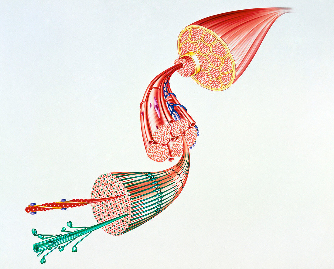
| Skeletal muscle. Illustration of the structure of human skeletal muscle. Skeletal muscle (top right) consists of bundles of long,thin cells called muscle fibres. A group of five muscle fibres with surrounding blood vessels is shown at centre. Each muscle fibre contains numerous "myofibrils",one of which is shown at lower centre. The myofibril is a bundle of two types of protein "myofilament": myosin (green) and actin (red). When the muscle is stimulated,tiny cross-bridges (lower left) in the myosin filaments hook onto actin filaments and pull them along. As a result,myosin and actin filaments slide over each other,shortening the muscle and making it contract | |
| Lizenzart: | Lizenzpflichtig |
| Credit: | Science Photo Library / Bavosi, John |
| Bildgröße: | 3543 px × 2853 px |
| Modell-Rechte: | nicht erforderlich |
| Eigentums-Rechte: | nicht erforderlich |
| Restrictions: | - |
Preise für dieses Bild ab 15 €
Universitäten & Organisationen
(Informationsmaterial Digital, Informationsmaterial Print, Lehrmaterial Digital etc.)
ab 15 €
Redaktionell
(Bücher, Bücher: Sach- und Fachliteratur, Digitale Medien (redaktionell) etc.)
ab 30 €
Werbung
(Anzeigen, Aussenwerbung, Digitale Medien, Fernsehwerbung, Karten, Werbemittel, Zeitschriften etc.)
ab 55 €
Handelsprodukte
(bedruckte Textilie, Kalender, Postkarte, Grußkarte, Verpackung etc.)
ab 75 €
Pauschalpreise
Rechtepakete für die unbeschränkte Bildnutzung in Print oder Online
ab 495 €