Pancreas
Bildnummer 11866544
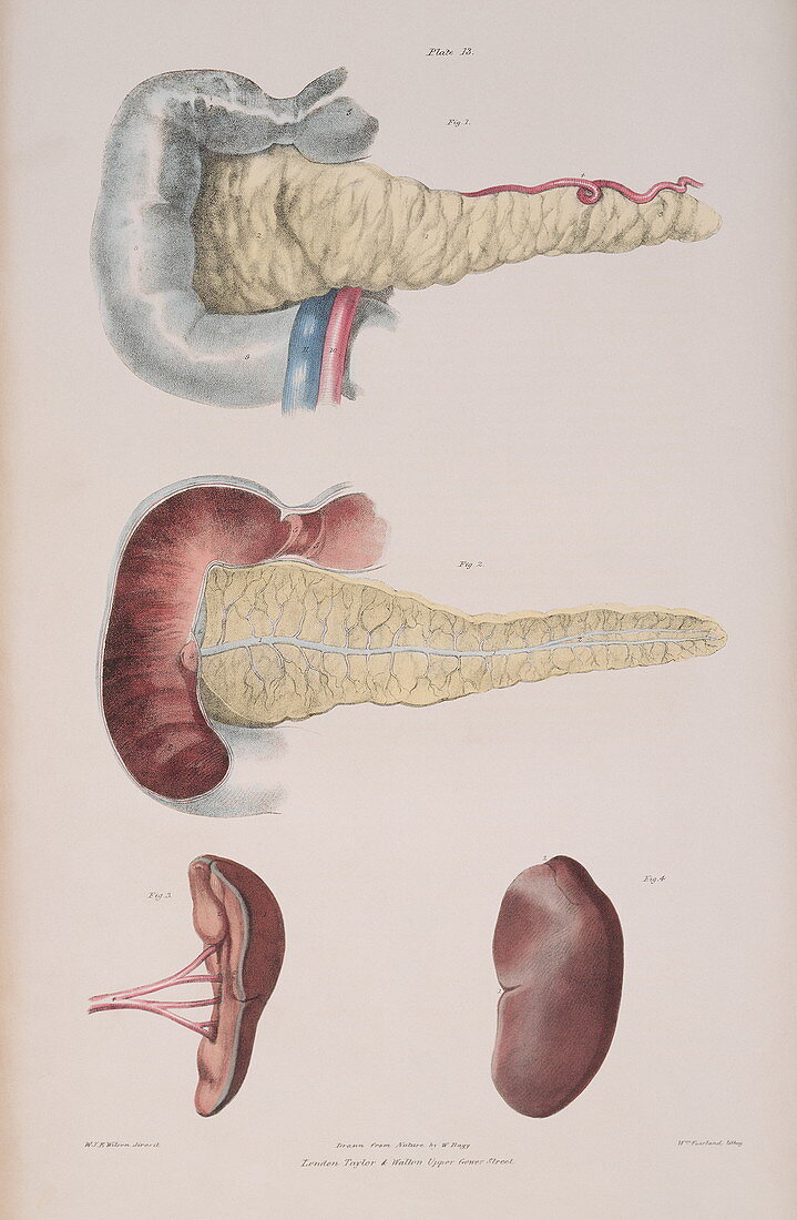
| Pancreas. Historical illustration of the pancreas (pale yellow) and the duodenum (pale blue,upper left). The centre diagram shows a cross-section through these organs. The ducts (pale blue) drain the digestive enzymes from the pancreas into the duodenum (here dark red),the first part of the small intestine. It is here that digestion of food from the stomach begins. The spleen is shown in the bottom two diagrams with the arterial blood supply shown at lower left. This organ contains lymphoid tissue,acts as a reservoir for blood and produces white blood cells for the immune system. Colour lithograph by Fairland from The Viscera of the Human Body,1840. Based on drawings by Bagg | |
| Lizenzart: | Lizenzpflichtig |
| Credit: | Science Photo Library / Terry, Sheila |
| Bildgröße: | 3043 px × 4656 px |
| Modell-Rechte: | nicht erforderlich |
| Eigentums-Rechte: | nicht erforderlich |
| Restrictions: | - |
Preise für dieses Bild ab 15 €
Universitäten & Organisationen
(Informationsmaterial Digital, Informationsmaterial Print, Lehrmaterial Digital etc.)
ab 15 €
Redaktionell
(Bücher, Bücher: Sach- und Fachliteratur, Digitale Medien (redaktionell) etc.)
ab 30 €
Werbung
(Anzeigen, Aussenwerbung, Digitale Medien, Fernsehwerbung, Karten, Werbemittel, Zeitschriften etc.)
ab 55 €
Handelsprodukte
(bedruckte Textilie, Kalender, Postkarte, Grußkarte, Verpackung etc.)
ab 75 €
Pauschalpreise
Rechtepakete für die unbeschränkte Bildnutzung in Print oder Online
ab 495 €