Brain in REM sleep
Bildnummer 11863974
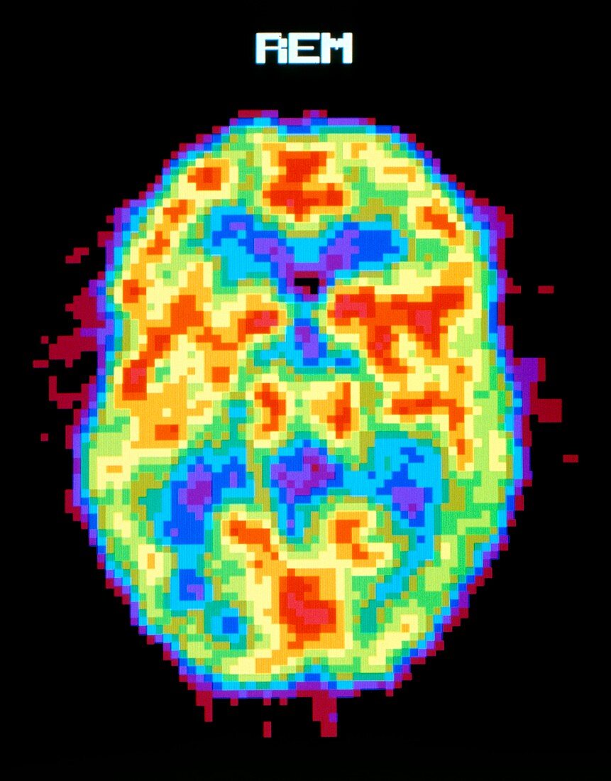
| Brain during REM sleep. Coloured Positron Emission Tomography (PET) scan of the human brain during REM (rapid eye movement) sleep. Colour-coding depicts active cerebral brain areas (red) through to inactive areas (blue). During the REM sleep phase,the brain is active and dreaming,showing similar activity when awake. In the non-REM phase of sleep the brain is in a deeper,less active sleep. PET scanning shows metabolic activity of the brain. A radioactive tracer (here,radio- labelled glucose) is injected into the bloodstream and absorbed by active tissues of the brain. The PET scanner detects photons emitted by the tracer,to produce a "slice" image of the brain | |
| Lizenzart: | Lizenzpflichtig |
| Credit: | Science Photo Library / Morgan, Hank |
| Bildgröße: | 3736 px × 4772 px |
| Modell-Rechte: | nicht erforderlich |
| Eigentums-Rechte: | nicht erforderlich |
| Restrictions: |
|
Preise für dieses Bild ab 15 €
Universitäten & Organisationen
(Informationsmaterial Digital, Informationsmaterial Print, Lehrmaterial Digital etc.)
ab 15 €
Redaktionell
(Bücher, Bücher: Sach- und Fachliteratur, Digitale Medien (redaktionell) etc.)
ab 30 €
Werbung
(Anzeigen, Aussenwerbung, Digitale Medien, Fernsehwerbung, Karten, Werbemittel, Zeitschriften etc.)
ab 55 €
Handelsprodukte
(bedruckte Textilie, Kalender, Postkarte, Grußkarte, Verpackung etc.)
ab 75 €
Pauschalpreise
Rechtepakete für die unbeschränkte Bildnutzung in Print oder Online
ab 495 €