Intervertebral bone graft,X-ray
Bildnummer 11855900
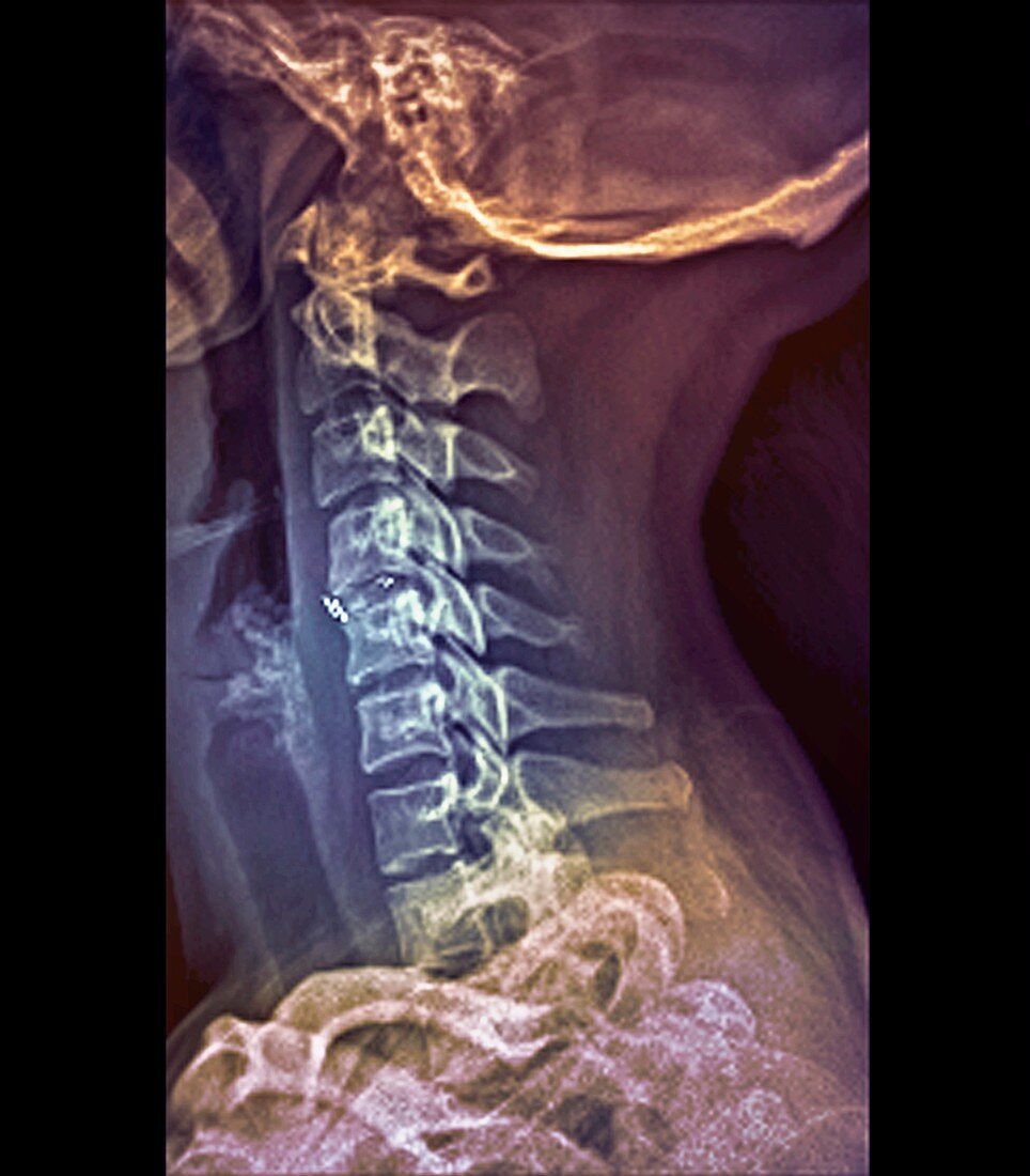
| Intervertebral bone graft. Coloured profile X-ray of the cervical vertebrae (neck bones) of a 29- year-old woman. The base of the skull is at top. A bone graft has been implanted in the intervertebral space (centre left) between the 5th and 6th cervical vertebrae (C5 and C6) in order to replace the intervertebral disc that has worn away. Intervertebral discs are flat,circular structures that contain cartilage and line the joints between the vertebrae. They act as shock absorbers and cushion the spine during movement | |
| Lizenzart: | Lizenzpflichtig |
| Credit: | Science Photo Library / Zephyr |
| Bildgröße: | 3458 px × 3946 px |
| Modell-Rechte: | nicht erforderlich |
| Eigentums-Rechte: | nicht erforderlich |
| Restrictions: | - |
Preise für dieses Bild ab 15 €
Universitäten & Organisationen
(Informationsmaterial Digital, Informationsmaterial Print, Lehrmaterial Digital etc.)
ab 15 €
Redaktionell
(Bücher, Bücher: Sach- und Fachliteratur, Digitale Medien (redaktionell) etc.)
ab 30 €
Werbung
(Anzeigen, Aussenwerbung, Digitale Medien, Fernsehwerbung, Karten, Werbemittel, Zeitschriften etc.)
ab 55 €
Handelsprodukte
(bedruckte Textilie, Kalender, Postkarte, Grußkarte, Verpackung etc.)
ab 75 €
Pauschalpreise
Rechtepakete für die unbeschränkte Bildnutzung in Print oder Online
ab 495 €
Keywords
- 20er Jahre,
- abnormal,
- Bandscheiben,
- behandelt,
- Behandlung,
- C5,
- C6,
- chirurgisch,
- eingefärbt,
- Erwachsene,
- farbig,
- Frau,
- geduldig,
- gefärbt,
- gepfropft,
- Gesundheitswesen,
- Hals,
- Halswirbel,
- Implantat,
- implantiert,
- Jung,
- Knochen,
- Medizin,
- medizinisch,
- menschlicher Körper,
- Operation,
- Osteologie,
- Radiographie,
- Reparatur,
- repariert,
- Röntgen,
- Röntgengerät,
- Transplantat,
- ungesund,
- vertebral,
- Weiblich,
- Wirbel,
- Wirbelsäule,
- zwanziger Jahre