Pregnancy ultrasound
Bildnummer 11848777
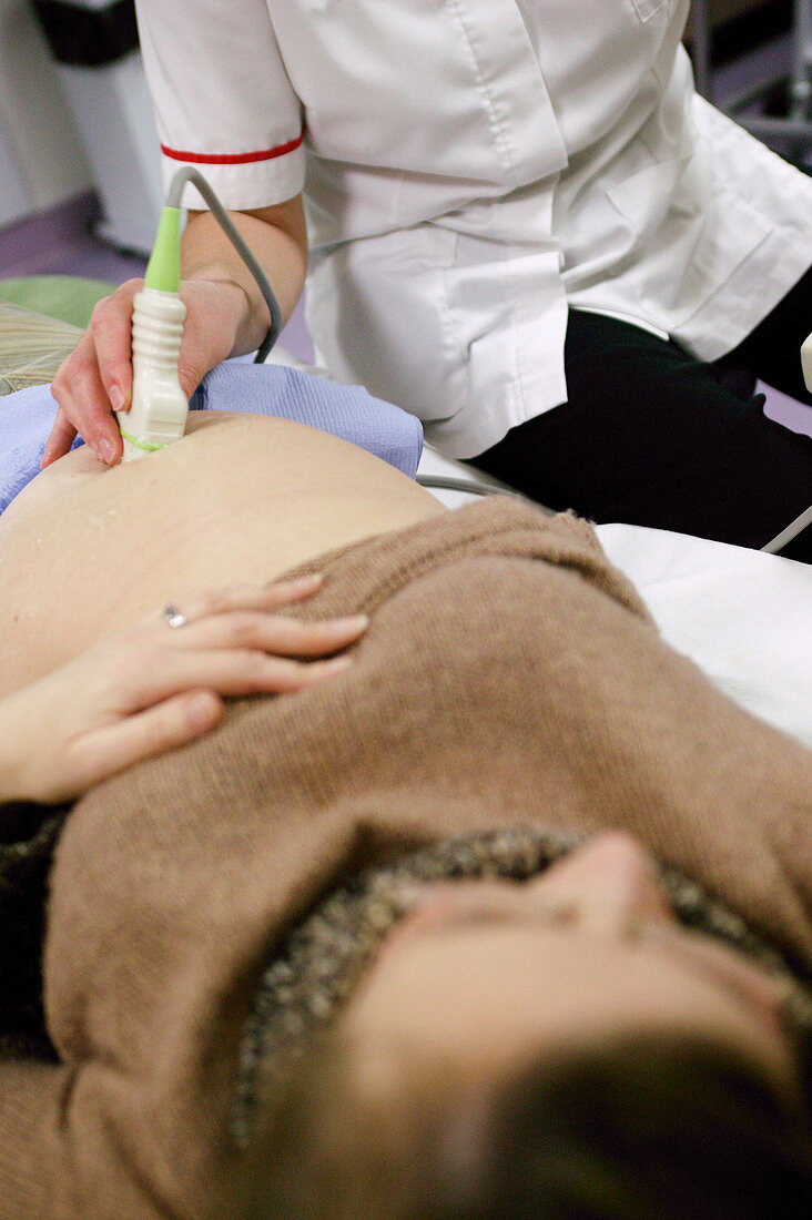
| Pregnancy ultrasound. Transducer (in hand) being used for an ultrasound scan of the swollen abdomen of a pregnant woman. The transducer emits high- frequency sound waves and detects the reflected echoes. The results are used to build an image (not seen) of the unborn child in the womb. This safe,non-invasive procedure is used routinely in pregnancy to assess the growth and health of the developing foetus,and to detect any abnormalities. This is a standard anomaly scan,carried out when the mother is 20 weeks pregnant. The detection of a serious anomaly at this stage (halfway through pregnancy),allows surgery or a termination to be considered | |
| Lizenzart: | Lizenzpflichtig |
| Credit: | Science Photo Library / Donne, Michael |
| Bildgröße: | 2704 px × 4064 px |
| Modell-Rechte: | nicht erforderlich |
| Eigentums-Rechte: | nicht erforderlich |
| Restrictions: | - |
Preise für dieses Bild ab 15 €
Universitäten & Organisationen
(Informationsmaterial Digital, Informationsmaterial Print, Lehrmaterial Digital etc.)
ab 15 €
Redaktionell
(Bücher, Bücher: Sach- und Fachliteratur, Digitale Medien (redaktionell) etc.)
ab 30 €
Werbung
(Anzeigen, Aussenwerbung, Digitale Medien, Fernsehwerbung, Karten, Werbemittel, Zeitschriften etc.)
ab 55 €
Handelsprodukte
(bedruckte Textilie, Kalender, Postkarte, Grußkarte, Verpackung etc.)
ab 75 €
Pauschalpreise
Rechtepakete für die unbeschränkte Bildnutzung in Print oder Online
ab 495 €
Keywords
- Abdomen,
- Arbeiter,
- Ausrüstung,
- Baby-Scan,
- Begutachten,
- Diagnose,
- fötal,
- Fötus,
- Frau,
- Geburtshelfer,
- Geburtshilfe,
- geburtshilflich,
- geduldig,
- gesund,
- Gesundheitswesen,
- Kontrolle,
- Krankenhaus,
- Krankenschwester,
- Maschine,
- Medizin,
- medizinisch,
- Menschen Person Personen,
- normal,
- Reproduktion,
- reproduktiv,
- Scan,
- Scanner,
- schwanger,
- Schwangerschaft,
- Sonogramm,
- Sonograph,
- Sonographie,
- Technik,
- Technologie,
- Untersuchung,
- Uterus,
- Weiblich,
- zwanzig