Malaria parasite,TEM
Bildnummer 11841045
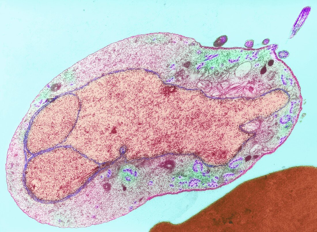
| Malaria parasite. Image 4 of 10. Coloured transmission electron micrograph (TEM) of a sexual male malaria (Plasmodium sp.) microgametocyte in a mosquito (Anopheles sp.) gut. The microgametocyte releases male microgametes (purple,one at top right) after it is ingested by a mosquito feeding on an infected human. Each motile gamete possesses a flagellum built from fibrils (purple,paired) assembled near the gametocyte membrane. The male gametes fertilise the female macrogametocytes (not seen). Asexual reproduction then produces the stage that infects humans. Magnification: x5700 at 6x7cm size. For the malaria life cycle,see images M210/197-206 | |
| Lizenzart: | Lizenzpflichtig |
| Credit: | Science Photo Library / Lshtm |
| Bildgröße: | 3500 px × 2560 px |
| Modell-Rechte: | nicht erforderlich |
| Eigentums-Rechte: | nicht erforderlich |
| Restrictions: | - |
Preise für dieses Bild ab 15 €
Universitäten & Organisationen
(Informationsmaterial Digital, Informationsmaterial Print, Lehrmaterial Digital etc.)
ab 15 €
Redaktionell
(Bücher, Bücher: Sach- und Fachliteratur, Digitale Medien (redaktionell) etc.)
ab 30 €
Werbung
(Anzeigen, Aussenwerbung, Digitale Medien, Fernsehwerbung, Karten, Werbemittel, Zeitschriften etc.)
ab 55 €
Handelsprodukte
(bedruckte Textilie, Kalender, Postkarte, Grußkarte, Verpackung etc.)
ab 75 €
Pauschalpreise
Rechtepakete für die unbeschränkte Bildnutzung in Print oder Online
ab 495 €
Keywords
- ansteckend,
- Assembler-,
- Bildung,
- Biologie,
- Bühne,
- elektronenmikroskopische Aufnahme,
- Erreger,
- Exflagellation,
- farbig,
- Formation,
- Gameten,
- Gewebe,
- horizontal,
- Infektion,
- infiziert,
- Insekt,
- Keimzelle,
- Krankheit,
- Krankheitserreger,
- Lebenszyklus,
- Mikrobe,
- Mikrobiologie,
- Mikrogameten,
- Mikroorganismus,
- Moskito,
- Parasit,
- parasitär,
- Parasiten,
- pathogen,
- Plasmodium sp.,
- Produktion,
- Protozoen,
- Protozoon,
- Reproduktion,
- Sektion,
- sektioniert,
- sexuell,
- tem,
- Übertragung