CT brain scan showing stroke (or CVA)
Bildnummer 11838221
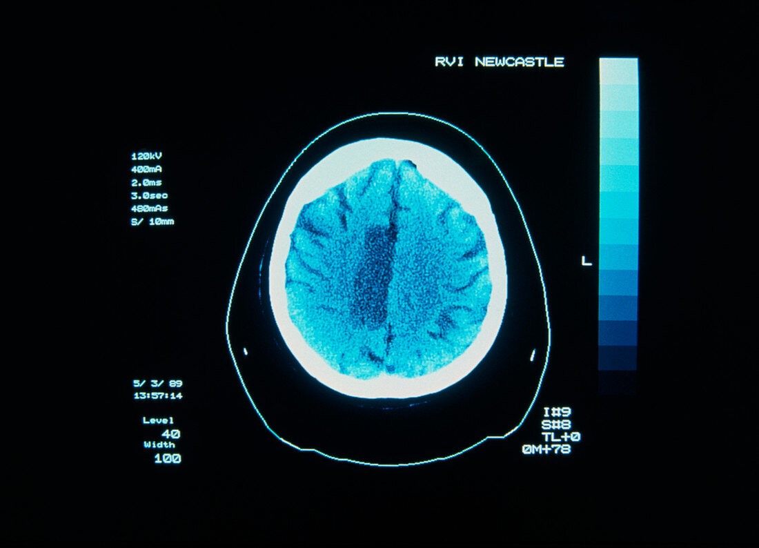
| Stroke: computed X-ray tomography (CT) scan of the brain of woman aged 48 years,revealing an area of cerebral infarction - the result of a stroke. The infarct - brain tissue starved of blood - appears as the dark rectangular area in centre. In CT,a narrow X-ray beam is directed through the subject towards a diametrically-opposed detector. A series of "slices" is made,with source & detector moving synchronously around the subject. Measurements of transmitted X-rays are processed by computer to reveal how elements of tissue in each "slice" affect the passage of X-rays & so construct an image of the section | |
| Lizenzart: | Lizenzpflichtig |
| Credit: | Science Photo Library / RVI, NEWCASTLE / SIMON FRASER |
| Bildgröße: | 5130 px × 3705 px |
| Modell-Rechte: | nicht erforderlich |
| Eigentums-Rechte: | nicht erforderlich |
| Restrictions: | - |
Preise für dieses Bild ab 15 €
Universitäten & Organisationen
(Informationsmaterial Digital, Informationsmaterial Print, Lehrmaterial Digital etc.)
ab 15 €
Redaktionell
(Bücher, Bücher: Sach- und Fachliteratur, Digitale Medien (redaktionell) etc.)
ab 30 €
Werbung
(Anzeigen, Aussenwerbung, Digitale Medien, Fernsehwerbung, Karten, Werbemittel, Zeitschriften etc.)
ab 55 €
Handelsprodukte
(bedruckte Textilie, Kalender, Postkarte, Grußkarte, Verpackung etc.)
ab 75 €
Pauschalpreise
Rechtepakete für die unbeschränkte Bildnutzung in Print oder Online
ab 495 €