3-D MRI scan of a brain with tumour
Bildnummer 11837871
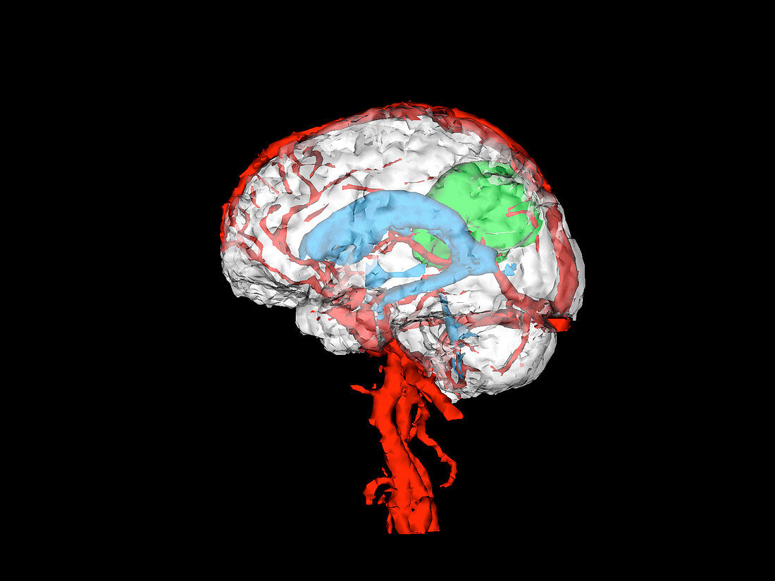
| Brain tumour. Coloured 3-D magnetic resonance imaging (MRI) scan of a patient's brain showing a brain tumour (green). Here,the blood vessels are red and the brain ventricle is cyan. This 3-D image was created from a series of MRI slice images through the brain. It will be used in 3-D virtual reality assisted surgery to help plan and execute the removal of the tumour. The image will be superimposed onto a video image of the patient's head. This allows the surgeon to locate the brain tumour in relation to other important tissues before surgery begins. The tumour can thus be removed with minimum damage to the brain | |
| Lizenzart: | Lizenzpflichtig |
| Credit: | Science Photo Library / BRIGHAM & WOMEN'S HOSPITAL / SURGICAL PLANNING LAB / MIT AI LAB |
| Bildgröße: | 3543 px × 2657 px |
| Modell-Rechte: | nicht erforderlich |
| Eigentums-Rechte: | nicht erforderlich |
| Restrictions: | - |
Preise für dieses Bild ab 15 €
Universitäten & Organisationen
(Informationsmaterial Digital, Informationsmaterial Print, Lehrmaterial Digital etc.)
ab 15 €
Redaktionell
(Bücher, Bücher: Sach- und Fachliteratur, Digitale Medien (redaktionell) etc.)
ab 30 €
Werbung
(Anzeigen, Aussenwerbung, Digitale Medien, Fernsehwerbung, Karten, Werbemittel, Zeitschriften etc.)
ab 55 €
Handelsprodukte
(bedruckte Textilie, Kalender, Postkarte, Grußkarte, Verpackung etc.)
ab 75 €
Pauschalpreise
Rechtepakete für die unbeschränkte Bildnutzung in Print oder Online
ab 495 €