PET brain scan: Alzheimer's disease
Bildnummer 11834394
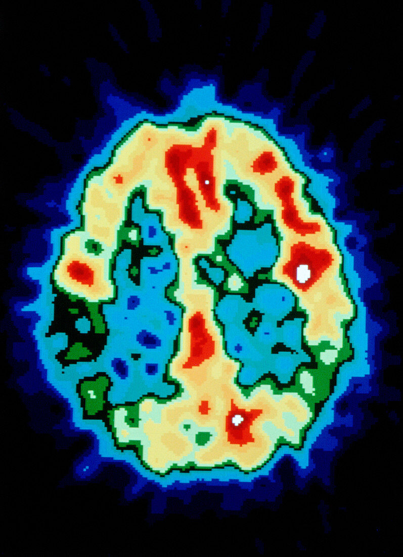
| Positron emission tomography (PET) scan of the brain of a patient with Alzheimer's disease (senile dementia). The colour-coded scan shows metabolic activity throughout this axial section,from low (blue) to high (red). PET scanning utilises an injected radioactive tracer (in this case an analogue of glucose) to reveal variations in metabolic activity in the brain. A normal scan would reveal a more symmetrical pattern of high activity across the cerebral hemispheres (the outer left & right areas of the image). Here,the blue-green area at bottom left reveals a particularly low level of metabolic activity. Alzheimer's is the most common form of dementia | |
| Lizenzart: | Lizenzpflichtig |
| Credit: | Science Photo Library / Pouedras, Catherine |
| Bildgröße: | 3157 px × 4356 px |
| Modell-Rechte: | nicht erforderlich |
| Eigentums-Rechte: | nicht erforderlich |
| Restrictions: |
|
Preise für dieses Bild ab 15 €
Universitäten & Organisationen
(Informationsmaterial Digital, Informationsmaterial Print, Lehrmaterial Digital etc.)
ab 15 €
Redaktionell
(Bücher, Bücher: Sach- und Fachliteratur, Digitale Medien (redaktionell) etc.)
ab 30 €
Werbung
(Anzeigen, Aussenwerbung, Digitale Medien, Fernsehwerbung, Karten, Werbemittel, Zeitschriften etc.)
ab 55 €
Handelsprodukte
(bedruckte Textilie, Kalender, Postkarte, Grußkarte, Verpackung etc.)
ab 75 €
Pauschalpreise
Rechtepakete für die unbeschränkte Bildnutzung in Print oder Online
ab 495 €