PET scan (basal ganglia) of Alzheimer's disease
Bildnummer 11834382
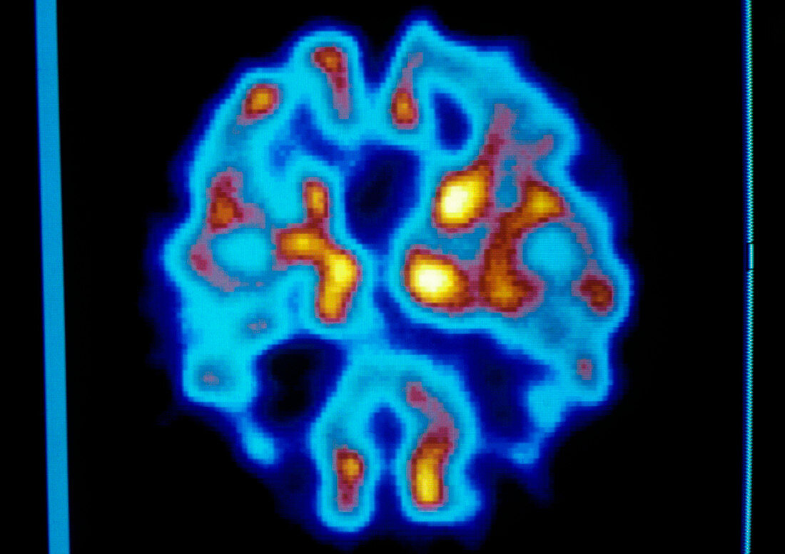
| Positron emission tomography (PET) scan of the brain (basal ganglia level) of a patient with senile dementia (Alzheimer's disease). The colour- coded scan through this cerebral layer shows brain activity: from low (blue) to high (yellow). PET scanning relies on an injected radioactive tracer to reveal variations in metabolic activity in the brain. Normal brain metabolic activity produces a roughly symmetrical pattern in the yellow areas of left and right cerebral hemispheres. The patchy appearance of this scan indicates degeneration of brain tissue. Symptoms of Alzheimer's disease include memory loss,personality changes,and disorientation. There is no known cure | |
| Lizenzart: | Lizenzpflichtig |
| Credit: | Science Photo Library / Beddow, Tim |
| Bildgröße: | 3560 px × 2516 px |
| Modell-Rechte: | nicht erforderlich |
| Eigentums-Rechte: | nicht erforderlich |
| Restrictions: | - |
Preise für dieses Bild ab 15 €
Universitäten & Organisationen
(Informationsmaterial Digital, Informationsmaterial Print, Lehrmaterial Digital etc.)
ab 15 €
Redaktionell
(Bücher, Bücher: Sach- und Fachliteratur, Digitale Medien (redaktionell) etc.)
ab 30 €
Werbung
(Anzeigen, Aussenwerbung, Digitale Medien, Fernsehwerbung, Karten, Werbemittel, Zeitschriften etc.)
ab 55 €
Handelsprodukte
(bedruckte Textilie, Kalender, Postkarte, Grußkarte, Verpackung etc.)
ab 75 €
Pauschalpreise
Rechtepakete für die unbeschränkte Bildnutzung in Print oder Online
ab 495 €