Degenerative disc disease,3D CT scan
Bildnummer 11763667
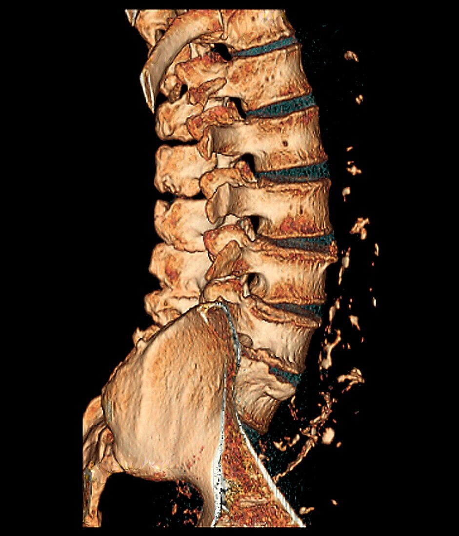
| Degenerative disc disease. Coloured 3D computed tomography (CT) scan of the lumbosacral spine of a 74 year old patient with degenerative disc disease. The disc between the S1 and L5 vertebrae (bottom) has completely degenerated and the disc between L5 and L4 (second from bottom) is partially degenerated. Calcified plaques are also seen in the aorta (down right) | |
| Lizenzart: | Lizenzfrei |
| Credit: | Science Photo Library / Zephyr |
| Modell-Rechte: | nicht erforderlich |
| Eigentums-Rechte: | nicht erforderlich |
| Restrictions: | - |
Preise für dieses Bild ab 29 €
Für digitale Nutzung (72 dpi)
ab 29 €
Für Druckauflösung (300 dpi)
ab 300 €
Keywords
- 3D,
- Anatomie,
- Aorta,
- ärztliche Untersuchung,
- Beschädigt,
- Biologie,
- CT-Scan,
- degenerativ,
- Dreidimensional,
- eine Person,
- Gesundheitswesen,
- Knochen,
- Knochenkrankheit,
- Krankheit,
- Kreislauf,
- Lendenwirbelsäule,
- Medizin,
- menschliche Anatomie,
- menschliches Körperteil,
- Plague,
- Radiographie,
- Röntgen,
- Rücken,
- Rückgrat,
- schwarzer Hintergrund,
- Skelett-,
- Struktur,
- unterer Rücken,
- Wirbel,
- Wirbelsäule