Dividing cancer cell,SEM
Bildnummer 11715590
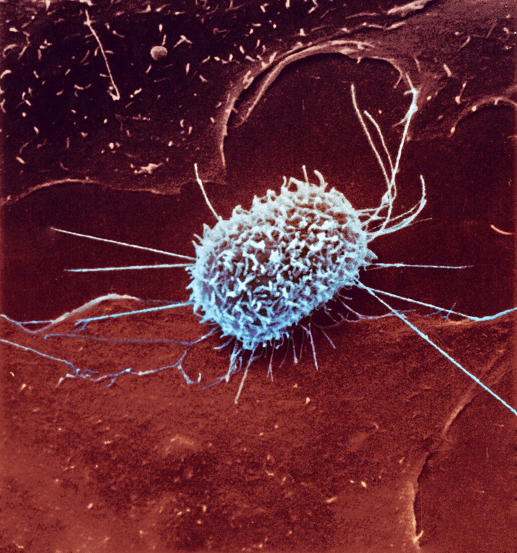
| Dividing cancer cell. Coloured scanning electron micrograph (SEM) of a cultured cancerous (malignant) cell from ovary tissue,showing the microvilli (finger-like projections) covering its surface. This surface appearance is typical,but not definitive,for cultured cells during the anaphase stage of mitotic cell division | |
| Lizenzart: | Lizenzpflichtig |
| Credit: | Science Photo Library / National Cancer Institute / AMI IMAGES |
| Bildgröße: | 3137 px × 3354 px |
| Modell-Rechte: | nicht erforderlich |
| Eigentums-Rechte: | nicht erforderlich |
| Restrictions: | - |
Preise für dieses Bild ab 15 €
Universitäten & Organisationen
(Informationsmaterial Digital, Informationsmaterial Print, Lehrmaterial Digital etc.)
ab 15 €
Redaktionell
(Bücher, Bücher: Sach- und Fachliteratur, Digitale Medien (redaktionell) etc.)
ab 30 €
Werbung
(Anzeigen, Aussenwerbung, Digitale Medien, Fernsehwerbung, Karten, Werbemittel, Zeitschriften etc.)
ab 55 €
Handelsprodukte
(bedruckte Textilie, Kalender, Postkarte, Grußkarte, Verpackung etc.)
ab 75 €
Pauschalpreise
Rechtepakete für die unbeschränkte Bildnutzung in Print oder Online
ab 495 €
Keywords
- abnormal,
- Eierstock,
- Eierstock-,
- einer,
- Farbig,
- gefärbt,
- Gesundheitswesen,
- Histopathologie,
- histopathologisch,
- Kondition,
- krank,
- Krankheit,
- Krebs,
- krebsartig,
- kultiviert,
- Kultur,
- maligne,
- Medizin,
- medizinisch,
- Mikrovilli,
- Mikrovillus,
- Mitose,
- Onkologie,
- onkologisch,
- Rasterelektronenmikroskop,
- rasterelektronenmikroskopische Aufnahme,
- REM,
- Säugetier,
- Säugetier-,
- Single,
- Störung,
- Teilen,
- ungesund,
- Zelle