Normal spine,3D CT scan
Bildnummer 11714190
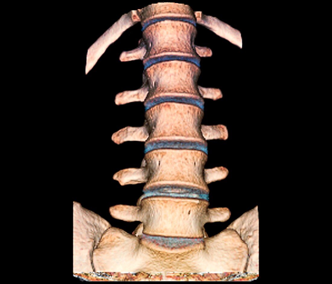
| Normal spine. Coloured 3D computed tomography (CT) scan of the rear of the healthy lumbar (lower back) spine of a 28-year-old,showing the normal structure of the lumbar vertebrae (L1-L5),and intervertebral discs,with the top of the pelvis (bottom) visible below. Each vertebra consists of the vertebral body and three processes,with two projecting out either side and one to the back | |
| Lizenzart: | Lizenzpflichtig |
| Credit: | Science Photo Library / Zephyr |
| Bildgröße: | 3854 px × 3307 px |
| Modell-Rechte: | nicht erforderlich |
| Eigentums-Rechte: | nicht erforderlich |
| Restrictions: | - |
Preise für dieses Bild ab 15 €
Universitäten & Organisationen
(Informationsmaterial Digital, Informationsmaterial Print, Lehrmaterial Digital etc.)
ab 15 €
Redaktionell
(Bücher, Bücher: Sach- und Fachliteratur, Digitale Medien (redaktionell) etc.)
ab 30 €
Werbung
(Anzeigen, Aussenwerbung, Digitale Medien, Fernsehwerbung, Karten, Werbemittel, Zeitschriften etc.)
ab 55 €
Handelsprodukte
(bedruckte Textilie, Kalender, Postkarte, Grußkarte, Verpackung etc.)
ab 75 €
Pauschalpreise
Rechtepakete für die unbeschränkte Bildnutzung in Print oder Online
ab 495 €
Keywords
- Anatomie,
- anatomisch,
- ausgeschnitten,
- Ausschnitte,
- Bandscheiben,
- Becken,
- diagnostische Bildgebung,
- farbig,
- Frontal,
- gefärbt,
- gesund,
- Hinter-,
- Knochen,
- L1,
- L2,
- L3,
- L4,
- L5,
- Lendenwirbelsäule,
- menschlicher Körper,
- Niemand,
- normal,
- pelvin,
- Prozess,
- Radiographie,
- Radiologie,
- radiologisch,
- Röntgen,
- Röntgenstrahlen,
- Röntgenstrahlung,
- Rückgrat,
- Scheibe,
- schwarzer Hintergrund,
- unterer Rücken,
- Wirbel,
- Wirbelsäule,
- Wirbelsäulen-