Fern (Adiantum capillus-veneris) leaf
Bildnummer 11712615
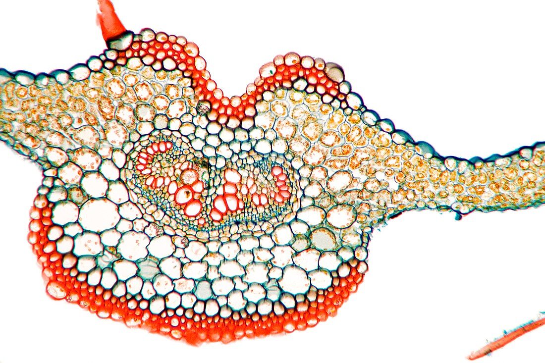
| Fern (Adiantum capillus-veneris) leaf. Light micrograph of a section through a leaf from a maidenhair fern,showing the midrib (vein,large,centre) containing the vascular bundle. Shown here are the upper epidermis (blue) and palisade mesophyll (green-yellow),containing chloroplasts. Beneath this are the parenchyma cells (yellow) and chloroplasts of the spongy mesophyll. The endodermis (brown) and inner pericycle (green). At centre is the vascular bundle consisting of metaxylem (red) and protoxylem (small cells,red),surrounding by the phloem sieve tube cells (green). Around the vascular bundle are parenchyma cells (large,blue). Magnification: x37 when printed 10 centimetres wide | |
| Lizenzart: | Lizenzpflichtig |
| Credit: | Science Photo Library / Wheeler, Dr. Keith |
| Bildgröße: | 5386 px × 3591 px |
| Modell-Rechte: | nicht erforderlich |
| Eigentums-Rechte: | nicht erforderlich |
| Restrictions: | - |
Preise für dieses Bild ab 15 €
Universitäten & Organisationen
(Informationsmaterial Digital, Informationsmaterial Print, Lehrmaterial Digital etc.)
ab 15 €
Redaktionell
(Bücher, Bücher: Sach- und Fachliteratur, Digitale Medien (redaktionell) etc.)
ab 30 €
Werbung
(Anzeigen, Aussenwerbung, Digitale Medien, Fernsehwerbung, Karten, Werbemittel, Zeitschriften etc.)
ab 55 €
Handelsprodukte
(bedruckte Textilie, Kalender, Postkarte, Grußkarte, Verpackung etc.)
ab 75 €
Pauschalpreise
Rechtepakete für die unbeschränkte Bildnutzung in Print oder Online
ab 495 €
Keywords
- Anatomie,
- anatomisch,
- Biologie,
- biologisch,
- Blatt,
- Botanik,
- botanisch,
- Chloroplasten,
- endodermal,
- Farn,
- Flora,
- Gewebe,
- Histologie,
- histologisch,
- Lichtmikroskop,
- lichtmikroskopische Aufnahme,
- Mittelrippe,
- Natur,
- Niemand,
- Parenchym,
- Pflanze,
- Pflanzen,
- Phloem,
- Röhren,
- Sektion,
- sektioniert,
- Tierwelt,
- vaskulär,
- Vene,
- Wedel,
- weißer Hintergrund,
- Xylem,
- Zelle,
- Zellen