Intervertebral disc,light micrograph
Bildnummer 11704726
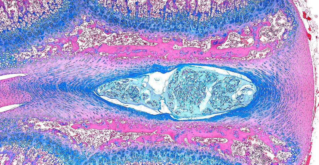
| Light microscopy of an intervertebral disc. The disc is found between two vertebral bodies their bone tissue margins stained pink. The centre of the disc is formed of cells and a gel-like matrix,called the nucleus pulposus. It is a remnant of the head-to-tail axis of the early embryo (the notochord). Fibrocartilage rings (blue) form the outer margins of the disc,called the nucleus fibrosus. The disc acts like a cushion between the stacked vertebral bodies and resists compression but allows movements of each vertebra. A slipped disc' is an abnormal protrusion (herniation) of the central portion through a damaged region or tear of the fibrocartilage. Magnification x60 when narrow width printed at 10 cm | |
| Lizenzart: | Lizenzpflichtig |
| Credit: | Science Photo Library / Microscape |
| Bildgröße: | 5851 px × 3034 px |
| Modell-Rechte: | nicht erforderlich |
| Eigentums-Rechte: | nicht erforderlich |
| Restrictions: | - |
Preise für dieses Bild ab 15 €
Universitäten & Organisationen
(Informationsmaterial Digital, Informationsmaterial Print, Lehrmaterial Digital etc.)
ab 15 €
Redaktionell
(Bücher, Bücher: Sach- und Fachliteratur, Digitale Medien (redaktionell) etc.)
ab 30 €
Werbung
(Anzeigen, Aussenwerbung, Digitale Medien, Fernsehwerbung, Karten, Werbemittel, Zeitschriften etc.)
ab 55 €
Handelsprodukte
(bedruckte Textilie, Kalender, Postkarte, Grußkarte, Verpackung etc.)
ab 75 €
Pauschalpreise
Rechtepakete für die unbeschränkte Bildnutzung in Print oder Online
ab 495 €