Skull in Erdheim-Chester disease,MRI
Bildnummer 11704664
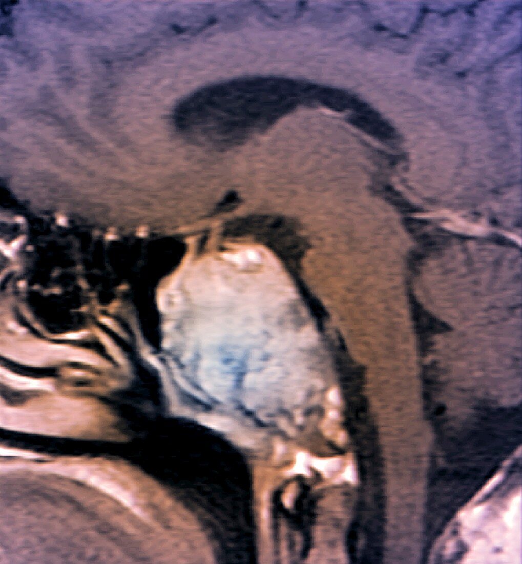
| Skull in Erdheim-Chester disease. Coloured computed tomography (CT) scan of an axial section through the head of a 28-old patient with Erdheim-Chester disease,showing osteosclerosis and bone erosion at the base of the skull,along with damage to the pituitary gland and pituitary stalk. Erdheim-Chester disease is the abnormal proliferation of histiocyte white blood cells,which causes osteosclerosis - increased bone density | |
| Lizenzart: | Lizenzpflichtig |
| Credit: | Science Photo Library / Zephyr |
| Bildgröße: | 2843 px × 3068 px |
| Modell-Rechte: | nicht erforderlich |
| Eigentums-Rechte: | nicht erforderlich |
| Restrictions: | - |
Preise für dieses Bild ab 15 €
Universitäten & Organisationen
(Informationsmaterial Digital, Informationsmaterial Print, Lehrmaterial Digital etc.)
ab 15 €
Redaktionell
(Bücher, Bücher: Sach- und Fachliteratur, Digitale Medien (redaktionell) etc.)
ab 30 €
Werbung
(Anzeigen, Aussenwerbung, Digitale Medien, Fernsehwerbung, Karten, Werbemittel, Zeitschriften etc.)
ab 55 €
Handelsprodukte
(bedruckte Textilie, Kalender, Postkarte, Grußkarte, Verpackung etc.)
ab 75 €
Pauschalpreise
Rechtepakete für die unbeschränkte Bildnutzung in Print oder Online
ab 495 €
Keywords
- abnormal,
- Abschnitte,
- Anatomie,
- anatomisch,
- Beschädigt,
- Computertomographie,
- ct,
- Degeneration,
- degenerativ,
- diagnostische Bildgebung,
- erodiert,
- Erosion,
- farbig,
- gefärbt,
- Gehirn,
- Gesundheitswesen,
- Kalzium,
- Knochen,
- Kondition,
- krank,
- Krankheit,
- Medizin,
- medizinisch,
- menschlicher Körper,
- Neuroimaging,
- Neurologie,
- neurologisch,
- Niemand,
- Osteologie,
- osteologisch,
- Radiographie,
- Radiologie,
- radiologisch,
- Röntgen,
- Röntgenstrahlen,
- Röntgenstrahlung,
- Scan,
- Schädel,
- Schaden,
- schwarzer Hintergrund,
- Sektion,
- sektioniert,
- Störung,
- ungesund,
- zentrales Nervensystem