Ear and cochlear anatomy,illustration
Bildnummer 11703752
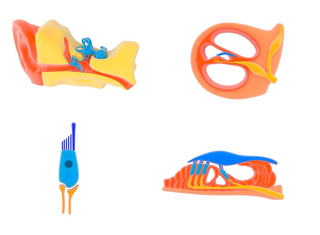
| Ear and cochlear anatomy. Illustration of the human ear (upper left) and successively expanded views of the anatomy of the cochlea,the organ of hearing in the inner ear. The cochlea is one of the organs shown in blue at upper left. At upper right,a sectioned view through the cochlea shows the cochlear duct with the vestibular canal and typanic canal either side. At lower right,the organ of Corti consists of the tectorial membrane (blue),hair cells (dark blue and light blue),and sensory neurons (orange) leading to the auditory nerve. A hair cell is at lower left,with its cilia and sensory microvilli (dark blue). For this artwork with labels,see C023/8843 | |
| Lizenzart: | Lizenzpflichtig |
| Credit: | Science Photo Library |
| Bildgröße: | 4922 px × 3621 px |
| Modell-Rechte: | nicht erforderlich |
| Eigentums-Rechte: | nicht erforderlich |
| Restrictions: | - |
Preise für dieses Bild ab 15 €
Universitäten & Organisationen
(Informationsmaterial Digital, Informationsmaterial Print, Lehrmaterial Digital etc.)
ab 15 €
Redaktionell
(Bücher, Bücher: Sach- und Fachliteratur, Digitale Medien (redaktionell) etc.)
ab 30 €
Werbung
(Anzeigen, Aussenwerbung, Digitale Medien, Fernsehwerbung, Karten, Werbemittel, Zeitschriften etc.)
ab 55 €
Handelsprodukte
(bedruckte Textilie, Kalender, Postkarte, Grußkarte, Verpackung etc.)
ab 75 €
Pauschalpreise
Rechtepakete für die unbeschränkte Bildnutzung in Print oder Online
ab 495 €
Keywords
- Amboss,
- Anatomie,
- anatomisch,
- aural,
- ausgeschnitten,
- Ausschnitte,
- Axone,
- Basilarmembran,
- Biochemie,
- biochemisch,
- Biologie,
- biologisch,
- Corti-Organ,
- Gehörgang,
- Gewebe,
- Hören,
- Illustration,
- Innenohr,
- Knochen,
- Kunstwerk,
- menschlicher Körper,
- Mikrovilli,
- Nerven,
- neural,
- Niemand,
- Ohr,
- Organ,
- Physiologie,
- physiologisch,
- sensorisch,
- Sinne,
- Tektorialmembran,
- weißer Hintergrund,
- Wimpern