Stomach cancer,barium X-ray
Bildnummer 11689378
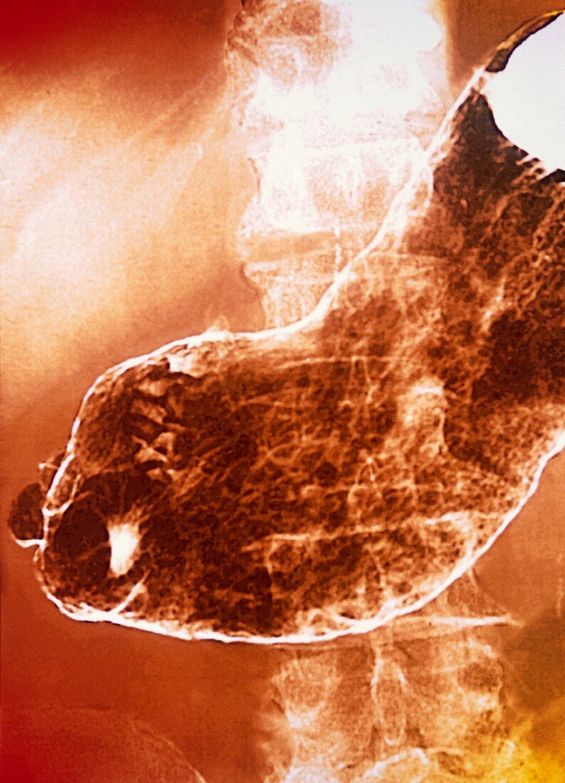
| Stomach cancer. Coloured frontal X-ray of a cancerous (malignant) tumour (dark,centre) in a patient's stomach. Sections of the gut show up as white because a radio-opaque barium meal was given prior to the X-ray. This highlights the structure of the gut. Any narrowing or obstructions,such as the cancer shown here,prevent the passage of the barium meal. They therefore do not show as white,allowing their location to be pinpointed | |
| Lizenzart: | Lizenzpflichtig |
| Credit: | Science Photo Library / GJLP |
| Bildgröße: | 3561 px × 4935 px |
| Modell-Rechte: | nicht erforderlich |
| Eigentums-Rechte: | nicht erforderlich |
| Restrictions: | - |
Preise für dieses Bild ab 15 €
Universitäten & Organisationen
(Informationsmaterial Digital, Informationsmaterial Print, Lehrmaterial Digital etc.)
ab 15 €
Redaktionell
(Bücher, Bücher: Sach- und Fachliteratur, Digitale Medien (redaktionell) etc.)
ab 30 €
Werbung
(Anzeigen, Aussenwerbung, Digitale Medien, Fernsehwerbung, Karten, Werbemittel, Zeitschriften etc.)
ab 55 €
Handelsprodukte
(bedruckte Textilie, Kalender, Postkarte, Grußkarte, Verpackung etc.)
ab 75 €
Pauschalpreise
Rechtepakete für die unbeschränkte Bildnutzung in Print oder Online
ab 495 €
Keywords
- Abdomen,
- abnormal,
- Bauch,
- Darm,
- Darm-,
- Diagnose,
- diagnostische Bildgebung,
- Eingeweide,
- farbig,
- Gastroenterologie,
- gastroenterologisch,
- Gedärme,
- gefärbt,
- GI tract,
- Kondition,
- Kontrastmittel,
- krank,
- Krankheit,
- Krebs,
- krebsartig,
- Magen-,
- Magen-Darm-Trakt,
- maligne,
- Malignom,
- Medizin,
- medizinisch,
- menschlicher Körper,
- Neoplasma,
- Onkologie,
- onkologisch,
- Radiographie,
- Radiologie,
- radiologisch,
- Röntgen,
- Röntgengerät,
- Röntgenstrahlen,
- Röntgenstrahlung,
- Störung,
- Tumor,
- ungesund,
- Verdauungssystem,
- Wachstum