Carpal tunnel wrist anatomy,artwork
Bildnummer 11678134
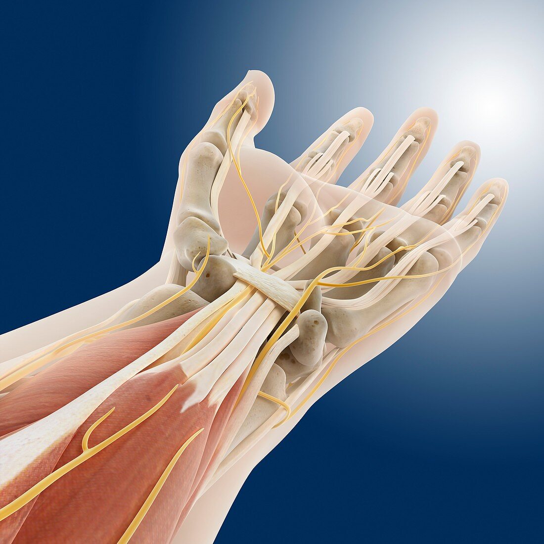
| Carpal tunnel wrist anatomy. Artwork of a palmar (palm-side up) view of the bones,tendons and ligaments (white),and nerves (yellow) of the hand and wrist,seen from the hand's proximal end. The skin surface is shown in outline,and some of the muscles (red) of the lower arm are also shown,with their tendons extending to the fingers. Clearly visible on the palmar side of the wrist is the anatomical structure called the carpal tunnel. Here,tendons and the median nerve pass through the annular ligament of the carpus. Repetitive overuse causing the median nerve to be compressed by the ligament is known as carpal tunnel syndrome | |
| Lizenzart: | Lizenzpflichtig |
| Credit: | Science Photo Library / Springer Medizin |
| Bildgröße: | 4180 px × 4180 px |
| Modell-Rechte: | nicht erforderlich |
| Eigentums-Rechte: | nicht erforderlich |
| Restrictions: | - |
Preise für dieses Bild ab 15 €
Universitäten & Organisationen
(Informationsmaterial Digital, Informationsmaterial Print, Lehrmaterial Digital etc.)
ab 15 €
Redaktionell
(Bücher, Bücher: Sach- und Fachliteratur, Digitale Medien (redaktionell) etc.)
ab 30 €
Werbung
(Anzeigen, Aussenwerbung, Digitale Medien, Fernsehwerbung, Karten, Werbemittel, Zeitschriften etc.)
ab 55 €
Handelsprodukte
(bedruckte Textilie, Kalender, Postkarte, Grußkarte, Verpackung etc.)
ab 75 €
Pauschalpreise
Rechtepakete für die unbeschränkte Bildnutzung in Print oder Online
ab 495 €