Prosthetic hip joint,artwork
Bildnummer 11651720
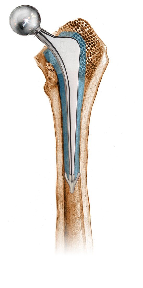
| Prosthetic hip joint. Cutaway artwork of a femur (thigh bone) showing a femoral component of a hip prosthesis. This component is implanted in the femur after the head of the femur has been surgically removed. The other components of the hip joint are a rounded ball (upper left) to fit into the socket implanted in the patient's pelvis (not shown). This allows the patient to regain mobility,and is usually done to treat severe osteoarthritis or a broken hip. This is an Exeter prosthesis,using bone cement (blue). Dating from 1970,it was developed by surgeons Robin Ling and Clive Lee in Exeter,UK | |
| Lizenzart: | Lizenzpflichtig |
| Credit: | Science Photo Library / D & L Graphics |
| Bildgröße: | 2648 px × 5315 px |
| Modell-Rechte: | nicht erforderlich |
| Eigentums-Rechte: | nicht erforderlich |
| Restrictions: | - |
Preise für dieses Bild ab 15 €
Universitäten & Organisationen
(Informationsmaterial Digital, Informationsmaterial Print, Lehrmaterial Digital etc.)
ab 15 €
Redaktionell
(Bücher, Bücher: Sach- und Fachliteratur, Digitale Medien (redaktionell) etc.)
ab 30 €
Werbung
(Anzeigen, Aussenwerbung, Digitale Medien, Fernsehwerbung, Karten, Werbemittel, Zeitschriften etc.)
ab 55 €
Handelsprodukte
(bedruckte Textilie, Kalender, Postkarte, Grußkarte, Verpackung etc.)
ab 75 €
Pauschalpreise
Rechtepakete für die unbeschränkte Bildnutzung in Print oder Online
ab 495 €
Keywords
- Arthritis,
- arthritisch,
- Arthrologie,
- Arthrose,
- ausgeschnitten,
- Ausrüstung,
- Ausschnitte,
- behandelt,
- Behandlung,
- Bein,
- beschriftet,
- britisch,
- chirurgisch,
- Cutaway,
- Design,
- Diagramm,
- einer,
- Englisch,
- Ersatz,
- Etikette,
- Etiketten,
- Exeter,
- Femur,
- Gelenk,
- Gerät,
- Hüfte,
- Illustration,
- implantiert,
- intern,
- Joint,
- Knochen,
- künstlich,
- Kunstwerk,
- Medizin,
- medizinisch,
- menschlicher Körper,
- Metall,
- Operation,
- Orthopädie,
- orthopädisch,
- Osteologie,
- osteologisch,
- Profil,
- Prothese,
- Reparatur,
- repariert,
- Sektion,
- sektioniert,
- Single,
- Technologie,
- technologisch,
- Vereinigtes Königreich,
- weißer Hintergrund,
- Welle