Prosthetic hip joint,diagram
Bildnummer 11651713
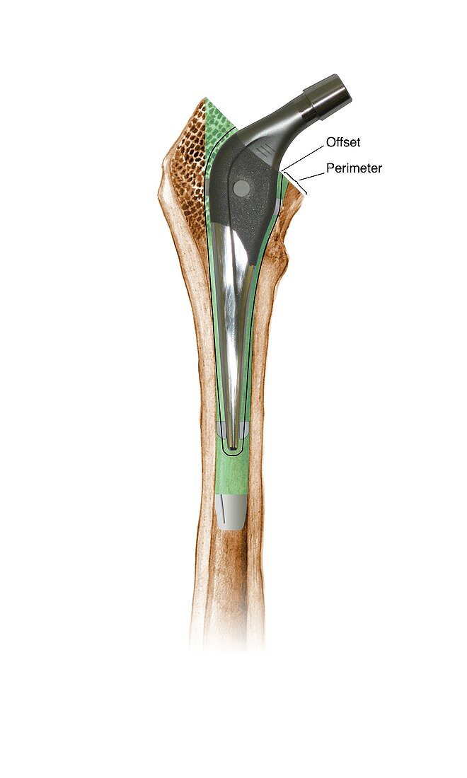
| Prosthetic hip joint. Cutaway diagram of a femur (thigh bone) showing a femoral component of a hip prosthesis. This component is implanted in the femur after the head of the femur has been surgically removed. The other components of the hip joint are a rounded ball to fit into the socket implanted in the patient's pelvis (not shown). This allows the patient to regain mobility,and is usually done to treat severe osteoarthritis or a broken hip. This is an SHP prosthesis,using bone cement (green). The 'offset' and 'perimeter' parameters are labelled. For this diagram with Gruen zones,see C016/6776 | |
| Lizenzart: | Lizenzpflichtig |
| Credit: | Science Photo Library / D & L Graphics |
| Bildgröße: | 3240 px × 5400 px |
| Modell-Rechte: | nicht erforderlich |
| Eigentums-Rechte: | nicht erforderlich |
| Restrictions: | - |
Preise für dieses Bild ab 15 €
Universitäten & Organisationen
(Informationsmaterial Digital, Informationsmaterial Print, Lehrmaterial Digital etc.)
ab 15 €
Redaktionell
(Bücher, Bücher: Sach- und Fachliteratur, Digitale Medien (redaktionell) etc.)
ab 30 €
Werbung
(Anzeigen, Aussenwerbung, Digitale Medien, Fernsehwerbung, Karten, Werbemittel, Zeitschriften etc.)
ab 55 €
Handelsprodukte
(bedruckte Textilie, Kalender, Postkarte, Grußkarte, Verpackung etc.)
ab 75 €
Pauschalpreise
Rechtepakete für die unbeschränkte Bildnutzung in Print oder Online
ab 495 €
Keywords
- Arthritis,
- arthritisch,
- Arthrologie,
- Arthrose,
- ausgeschnitten,
- Ausrüstung,
- Ausschnitte,
- behandelt,
- Behandlung,
- Bein,
- beschriftet,
- chirurgisch,
- Cutaway,
- Diagramm,
- einer,
- Ersatz,
- Etikette,
- Etiketten,
- Femur,
- Gelenk,
- Gerät,
- Hüfte,
- Illustration,
- implantiert,
- intern,
- Joint,
- Knochen,
- künstlich,
- Kunstwerk,
- Medizin,
- medizinisch,
- menschlicher Körper,
- Metall,
- Operation,
- Orthopädie,
- orthopädisch,
- Osteologie,
- osteologisch,
- Profil,
- Prothese,
- Reparatur,
- repariert,
- Sektion,
- sektioniert,
- Single,
- Technologie,
- technologisch,
- weißer Hintergrund,
- Welle