DNA bundle on silicon nanopillars,SEM
Bildnummer 11644074
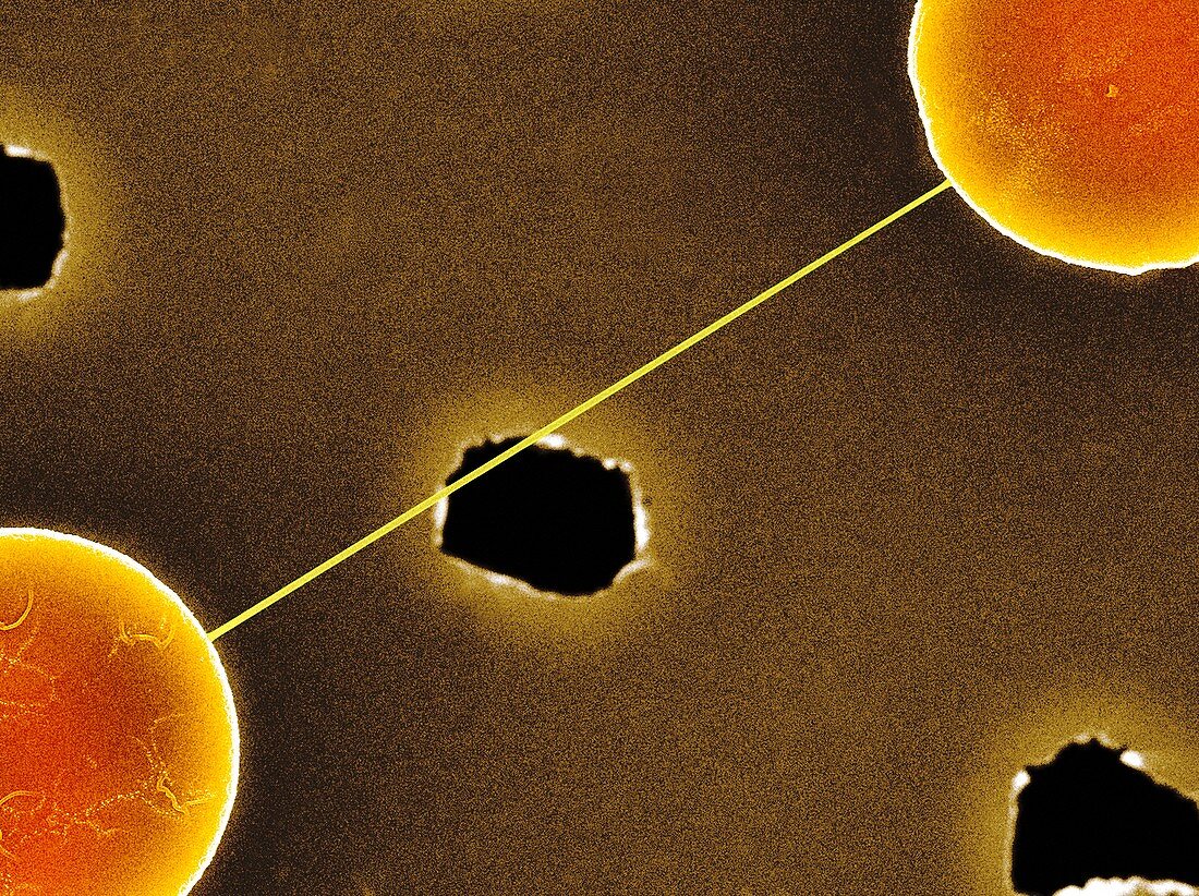
| DNA bundle on silicon nanopillars. Coloured scanning electron micrograph (SEM) of a DNA (deoxyribonucleic acid) bundle and silicon nanopillars used to obtain the first high-contrast direct images of DNA. The DNA bundle was prepared on a bed of silicon nanopillars (bottom left and top right). The holes allow the passage of electrons for transmission electron microscopy (TEM). The bundle (several DNA strands) is around 20 nanometres wide. This image was obtained in 2012 by a team led by Enzo di Fabrizio from the Department of Nanostructure,at the Italian Institute of Technology (IIT) in Genoa. For the TEM image,see C015/3216 | |
| Lizenzart: | Lizenzpflichtig |
| Credit: | Science Photo Library / PROFESSOR ENZO DI FABRIZIO, IIT |
| Bildgröße: | 4843 px × 3621 px |
| Modell-Rechte: | nicht erforderlich |
| Eigentums-Rechte: | nicht erforderlich |
| Restrictions: | - |
Preise für dieses Bild ab 15 €
Universitäten & Organisationen
(Informationsmaterial Digital, Informationsmaterial Print, Lehrmaterial Digital etc.)
ab 15 €
Redaktionell
(Bücher, Bücher: Sach- und Fachliteratur, Digitale Medien (redaktionell) etc.)
ab 30 €
Werbung
(Anzeigen, Aussenwerbung, Digitale Medien, Fernsehwerbung, Karten, Werbemittel, Zeitschriften etc.)
ab 55 €
Handelsprodukte
(bedruckte Textilie, Kalender, Postkarte, Grußkarte, Verpackung etc.)
ab 75 €
Pauschalpreise
Rechtepakete für die unbeschränkte Bildnutzung in Print oder Online
ab 495 €
Keywords
- 2012,
- 21. Jahrhundert,
- Abteilung für Nanostruktur,
- Ballaststoff,
- Biochemie,
- biochemisch,
- Biologie,
- biologisch,
- Biomolekül,
- Bündeln,
- Desoxiribonukleinsäure,
- DNA,
- Doppelhelix,
- Enzo Di Fabrizio,
- erst,
- europäisch,
- farbig,
- Forschung,
- gefärbt,
- Genetik,
- Geschichte,
- Helix,
- Historisch,
- Italienisch,
- Italienisches Institut für Technologie,
- Löcher,
- Makromolekül,
- Nukleinsäure,
- Pionier,
- Rasterelektronenmikroskop,
- rasterelektronenmikroskopische Aufnahme,
- REM,
- sprialförmig,
- Strand,
- strukturell,
- Transmissionselektronenmikroskopie,
- wegweisend