Fossil microtomography scanning
Bildnummer 11623781
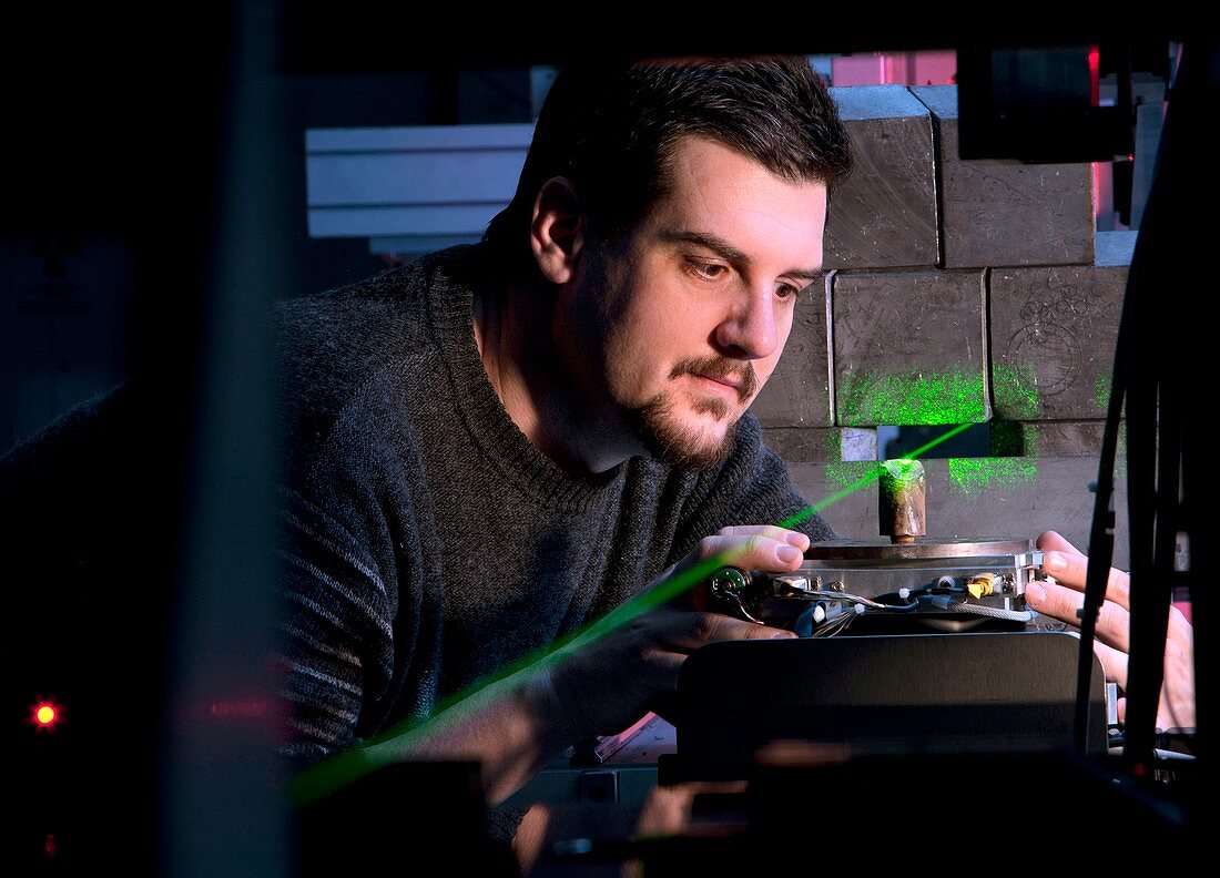
| Fossil microtomography scanning. Researcher positioning a Baculites fossil on a beamline (green) for a microtomography scan. This scanning technique uses high-resolution X-ray scanning (computed tomography) to record microscopic internal structure. An advanced version is synchrotron X-ray tomographic microscopy (SRXTM). This is the ID19 beamline at the European Synchrotron Radiation Facility (ESRF),in Grenoble,France. Baculites is a straight-shelled form of ammonite. The specimen is from the French National Museum of Natural History (MNHN),Paris,France. Photographed in 2011 | |
| Lizenzart: | Lizenzpflichtig |
| Credit: | Science Photo Library / Goetgheluck, Pascal |
| Bildgröße: | 3824 px × 2752 px |
| Modell-Rechte: | Derzeit liegt noch kein Release vor. Bitte kontaktieren Sie uns vor Verwendung. |
| Eigentums-Rechte: | nicht erforderlich |
| Restrictions: | - |
Preise für dieses Bild ab 15 €
Universitäten & Organisationen
(Informationsmaterial Digital, Informationsmaterial Print, Lehrmaterial Digital etc.)
ab 15 €
Redaktionell
(Bücher, Bücher: Sach- und Fachliteratur, Digitale Medien (redaktionell) etc.)
ab 30 €
Werbung
(Anzeigen, Aussenwerbung, Digitale Medien, Fernsehwerbung, Karten, Werbemittel, Zeitschriften etc.)
ab 55 €
Handelsprodukte
(bedruckte Textilie, Kalender, Postkarte, Grußkarte, Verpackung etc.)
ab 75 €
Pauschalpreise
Rechtepakete für die unbeschränkte Bildnutzung in Print oder Online
ab 495 €
Keywords
- 2011,
- 21. Jahrhundert,
- Ausrüstung,
- Baculites,
- Bernstein,
- Biologe,
- Biologie,
- biologisch,
- Erwachsene,
- ESRF,
- Europa,
- europäisch,
- Fauna,
- forschend,
- Forscher,
- Forschung,
- Fossil,
- Fossilien,
- fossilisiert,
- Frankreich,
- Französisch,
- Geschichte,
- Grenoble,
- historisch,
- Ingenieurwesen,
- kaukasisch,
- Labor,
- Laser,
- Mann,
- Männlich,
- Maschine,
- Mensch,
- Menschen,
- MNHN,
- Modelling,
- Molluske,
- Museum,
- Muster,
- Naturgeschichte,
- nicht-invasive Technik,
- Paläontologe,
- Paläontologie,
- paläontologisch,
- Paläozoologie,
- Paul Tafforeau,
- Person,
- prähistorisch,
- Probe,
- Scannen,
- Scanner,
- Technologie,
- technologisch,
- Tier,
- Tierwelt,
- versteinert,
- Vorgeschichte,
- weiß,
- wirbellos,
- Wissenschaftler,
- Zoologe,
- Zoologie,
- zoologisch