Normal human knee,artwork
Bildnummer 11609060
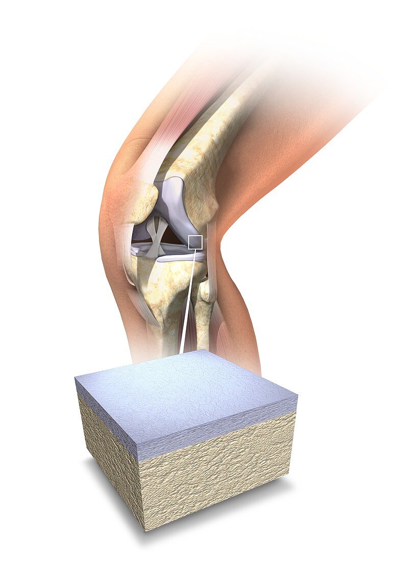
| Normal human knee. Artwork showing the internal anatomy of a healthy human knee,with an inset (bottom) of the cartilage (grey) that covers the ends of the bones forming this joint. The cartilage cushions the joint,ensuring smooth movement. The bones are the femur (thigh bone,upper right),the patella (knee-cap,left),and the lower leg bones the tibia (larger) and fibula (smaller). Several ligaments (white straps) hold the bones together. The patellar tendon (actually a ligament) joins the knee-cap to the tibia. The lateral collateral ligament joins the femur and fibula. The crossed cruciate ligaments join the femur and tibia | |
| Lizenzart: | Lizenzpflichtig |
| Credit: | Science Photo Library / Dalhoff, Henning |
| Bildgröße: | 3543 px × 4961 px |
| Modell-Rechte: | nicht erforderlich |
| Eigentums-Rechte: | nicht erforderlich |
| Restrictions: | - |
Preise für dieses Bild ab 15 €
Universitäten & Organisationen
(Informationsmaterial Digital, Informationsmaterial Print, Lehrmaterial Digital etc.)
ab 15 €
Redaktionell
(Bücher, Bücher: Sach- und Fachliteratur, Digitale Medien (redaktionell) etc.)
ab 30 €
Werbung
(Anzeigen, Aussenwerbung, Digitale Medien, Fernsehwerbung, Karten, Werbemittel, Zeitschriften etc.)
ab 55 €
Handelsprodukte
(bedruckte Textilie, Kalender, Postkarte, Grußkarte, Verpackung etc.)
ab 75 €
Pauschalpreise
Rechtepakete für die unbeschränkte Bildnutzung in Print oder Online
ab 495 €
Keywords
- ACL,
- Anatomie,
- anatomisch,
- Arthrologie,
- Artikulation,
- ausgeschnitten,
- Ausschnitte,
- Band,
- Bänder,
- Biologie,
- biologisch,
- Femur,
- Gelenk,
- gesund,
- Gewebe,
- hinteres Kreuzband,
- Illustration,
- Joint,
- Knie,
- Knochen,
- Knorpel,
- Kunstwerk,
- laterales Seitenband,
- menschlicher Körper,
- normal,
- Orthopädie,
- pcl,
- Schenkel,
- Schienbein,
- Unterschenkel,
- Wadenbein