Developing frog egg,light micrograph
Bildnummer 11593517
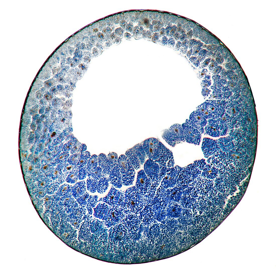
| Developing frog egg. Light micrograph of a vertical section through a developing egg laid by a common frog (Rana temporaria temporaria). The egg has reached the blastula stage. The cell cleavages of the egg are not equal so the animal pole (top) has more and smaller cells,whilst the yolk at the vegetative end (bottom) slows down cleavage and results in fewer and larger cells. The vitelline membrane surrounds the egg. Inside the blastula there is a hollow filled with fluid,the blastocoele. Magnification: x100 when printed at 10 centimetres across | |
| Lizenzart: | Lizenzpflichtig |
| Credit: | Science Photo Library / Wheeler, Dr. Keith |
| Bildgröße: | 4264 px × 4264 px |
| Modell-Rechte: | nicht erforderlich |
| Eigentums-Rechte: | nicht erforderlich |
| Restrictions: | - |
Preise für dieses Bild ab 15 €
Universitäten & Organisationen
(Informationsmaterial Digital, Informationsmaterial Print, Lehrmaterial Digital etc.)
ab 15 €
Redaktionell
(Bücher, Bücher: Sach- und Fachliteratur, Digitale Medien (redaktionell) etc.)
ab 30 €
Werbung
(Anzeigen, Aussenwerbung, Digitale Medien, Fernsehwerbung, Karten, Werbemittel, Zeitschriften etc.)
ab 55 €
Handelsprodukte
(bedruckte Textilie, Kalender, Postkarte, Grußkarte, Verpackung etc.)
ab 75 €
Pauschalpreise
Rechtepakete für die unbeschränkte Bildnutzung in Print oder Online
ab 495 €
Keywords
- ausgeschnitten,
- Ausschnitte,
- befleckt,
- Biologie,
- biologisch,
- Bühne,
- Ei,
- Embryo,
- Entwicklung,
- Fauna,
- Frosch,
- Gewebe,
- Herpetologie,
- Kreis,
- kreisförmig,
- Lichtmikroskop,
- lichtmikroskopische Aufnahme,
- Natur,
- Reproduktion,
- rund,
- Sektion,
- sektioniert,
- Stationen,
- Tier,
- Tierwelt,
- Verfärbung,
- Zelle,
- Zellen,
- zellular,
- Zoologie,
- zoologisch